2012 Group Project 6: Difference between revisions
| Line 71: | Line 71: | ||
The first evidence regarding the contribution of Wnt signalling came from experiments with the otic ectoderm of chicks. Data showed that specific marker genes, such as Pax2, were induced to a greater extend with FGF19 and Wnt8c present as compared to FGF19 alone <ref><pubmed>11110663</pubmed></ref>. Ladher et al.(2000) hypothesised that FGF19 induced Wnt8c, and together they induced the otic gene markers. | The first evidence regarding the contribution of Wnt signalling came from experiments with the otic ectoderm of chicks. Data showed that specific marker genes, such as Pax2, were induced to a greater extend with FGF19 and Wnt8c present as compared to FGF19 alone <ref><pubmed>11110663</pubmed></ref>. Ladher et al.(2000) hypothesised that FGF19 induced Wnt8c, and together they induced the otic gene markers. | ||
Another possibility is the independent action of FGFs and Wnt signalling. Wnt signalling suppresses Foxi2, resulting in a Foxi2-negative area of particular size, which then allows for FGFs to induce otic genes | Another possibility is the independent action of FGFs and Wnt signalling. Wnt signalling suppresses Foxi2, resulting in a Foxi2-negative area of particular size, which then allows for FGFs to induce otic genes <ref><pubmed>16452098</pubmed></ref>. In this case, Wnt signalling determined the size of the otic placode, yet acted independently from FGF signalling. | ||
==Abnormal Hearing== | ==Abnormal Hearing== | ||
Revision as of 10:13, 3 September 2012
Hearing Development
Introduction
History
Adult Anatomy and Histology
Outer ear: Pinna, Auricle and Tympanic membrane
Middle ear: Ossicles (Malleus, Incus and Stapes) and Muscles (Tensor Tympani and Stapedius)
Inner ear: Bony and Membranous Labyrinth - Cochlea containing the Organ of corti, Vestibule containing Utricle and Saccule and Semi-circular canals containing semi-circular ducts
Development
The development of outer ear is attributed to the first pharyngeal arch. All the germ layer namely endoderm, mesoderm and ectoderm contribute to its formation. [1]
The pharyngeal arches arise as a series of bulges arising laterally from the embryo head around 3-4 weeks of human development. The arches have a consistent organisation of the endoderm, ectoderm and mesoderm. The ectoderm forms the outer surface of the arch with the core made up of mesoderm. Next to the mesoderm core on the opposite the ectoderm is the endoderm. All three layers contribute to the formation of outer, middle and inner ear structures hence contributing to hearing. In between the arches the ectoderm and endoderm come in contact with each other forming a continuous sheath on either side of the mesoderm, forming groves externally and arches internally. Inside the mesoderm core of each arch lies a specific cell population that goes onto develop into a nerve, cartilage and artery.
Outer Ear
The pinna is derived from the first pharyngeal cleft from the many protuberances that occur between the first and second pharyngeal arches. These protuberances are called auricular hillocks each of which develops into a specific part of the pinna. [2]
The external acoustic meatus is derived from the pharyngeal groove located between the first and second pharyngeal arches or the mandibular and hyoid arches.[3]
Middle Ear
Inner Ear
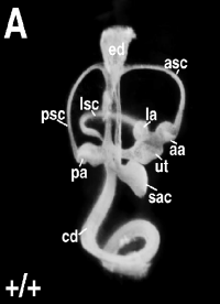
The entire inner ear, as well as the neurons which innervate the sensory organ, are derived from the otic placode. The otic placode is a thickened portion of ectoderm located on the each side of the developing head of the embryo, next to the hindbrain. It is generally visible after gastrulation, once the first 5 to 10 pairs of somites have formed. Invagination occurs next, which creates the otocyst - a vesicle which will develop into the different components of the inner ear: the cochlea, the semicircular canals with cristae, the utricle, the saccule and the vestibulo-acoustic ganglion. [4]
Induction of the otic placode
Experiments with molecular markers have revealed that several steps are needed for induction of the otic placode. We will briefly consider the three major steps:
1 Pre-placodal domain
The pre-placodal domain is a narrow strip of the ectoderm adjacent to the anterior neural plate after gastrulation. Different placodes arise from the pre-placodal domain. All the craniofacial sensory organs, including the ear, develop from these different placodes located at the periphery of the neural plate.
Various evidence indicates the existence of this pre-placodal region:
- Morphology indicates a thickened band of ectoderm around the anterior neural plate in some species, including mice and humans. As time progresses, this thickening will only be present at the locations where the different craniofacial placodes differentiate - including the otic placode.[5]
- Experiments have also shown that the placodes will only develop in the correct location, if rotation of the ectoderm along the anteroposterior axis takes place at the open neural plate stage. If rotation takes place at a later time, the placodes will form at incorrect places.[6]
- Gene expression has also indicated that particular genes are present in the pre-placodal domain. These genes belong to the Dlx, Six, Eya, Iro, BMP, Foxi and Msx families. Glavic et al. (2004) has shown that 'loss and gain of function of some of these genes resulted in the widening or reduction of the pre-placodal field'. Linked to this was also the domain of expression of some placode-specific genes; which either enlarged or diminished.[7]
2 Pre-otic field
Once the general placodal state has been established, the identity of each placode is induced by local signals. The optic placode is induced by various signals, including Pax8, Pax2, Fibroblast Growth Factors (FGFs), and many transciption factors.
In particular the FGFs are significant otic inducers. Signalling occurs from various rhombomeres from the hindbrain and the cranial paraxial mesoderm located beneath the area of the otic placode. For example, in mice FGF3 is expressed in rhombomeres 5 and 6, whereas FGF10 is expressed in the underlying mesoderm.[8] [9] Mutations of FGF3 and FGF10 have been investigated. Results showed that mice with a mutation of either FGF3 or FGF10 developed an abnormal otic vesicle, and a combination of the two mutants resulted in failure to form an otic vesicle.[9]
--> picture
3 Otic placode/epidermis fate decision
In the presence of FGF signalling, Wnt signalling can significantly influence the next step, which is the otic placode/epidermis fate decision. According to […] ‘Cells cells receiving high levels of Wnt signaling differentiating as otic placode, while cells receiving little or no Wnt signaling differentiating as epidermis.’
The first evidence regarding the contribution of Wnt signalling came from experiments with the otic ectoderm of chicks. Data showed that specific marker genes, such as Pax2, were induced to a greater extend with FGF19 and Wnt8c present as compared to FGF19 alone [10]. Ladher et al.(2000) hypothesised that FGF19 induced Wnt8c, and together they induced the otic gene markers.
Another possibility is the independent action of FGFs and Wnt signalling. Wnt signalling suppresses Foxi2, resulting in a Foxi2-negative area of particular size, which then allows for FGFs to induce otic genes [11]. In this case, Wnt signalling determined the size of the otic placode, yet acted independently from FGF signalling.
Abnormal Hearing
Genetic
Syndromic hearing loss
Non syndromic hearing loss
1 Mutation of GJB2 gene
Environmental
Infections
1 Rubella
2 Herpes
3 Syphilis
4 Toxoplasmosis
Drugs
1 Alcohol consumption during pregnancy
2 Chemotherapy
3 Accutane
Structural malformation of the ear
1 Stenosis
2 Enlarged vestibular aqueduct
3 Auricular Appendages
4 Absence of the Auricle
5 Microtia
6 Preauricular Sinuses and fistulas
7 Atresia of External Acoustic Meatus
8Absence of external acoustic meatus
9 Congenital cholesteatoma
Technologies to detect
Technologies to overcome the problems
Current Research
Glossary
References
- ↑ <pubmed>8287791</pubmed>
- ↑ <pubmed>14674478</pubmed>
- ↑ <pubmed>10976045</pubmed>
- ↑ <pubmed>17891709</pubmed>
- ↑ <pubmed>15531360</pubmed>
- ↑ <pubmed>14100031</pubmed>
- ↑ <pubmed>15242793</pubmed>
- ↑ <pubmed>7789270</pubmed>
- ↑ 9.0 9.1 <pubmed>12810586</pubmed>
- ↑ <pubmed>11110663</pubmed>
- ↑ <pubmed>16452098</pubmed>
External Links
External Links Notice - The dynamic nature of the internet may mean that some of these listed links may no longer function. If the link no longer works search the web with the link text or name. Links to any external commercial sites are provided for information purposes only and should never be considered an endorsement. UNSW Embryology is provided as an educational resource with no clinical information or commercial affiliation.
--Mark Hill 12:22, 15 August 2012 (EST) Please leave the content listed below the line at the bottom of your project page.
2012 Projects: Vision | Somatosensory | Taste | Olfaction | Abnormal Vision | Hearing
