File:Mouse oocytes in vitro.png
From Embryology

Size of this preview: 800 × 577 pixels. Other resolutions: 2,560 × 1,847 pixels | 3,439 × 2,481 pixels.
Original file (3,439 × 2,481 pixels, file size: 4.19 MB, MIME type: image/png)
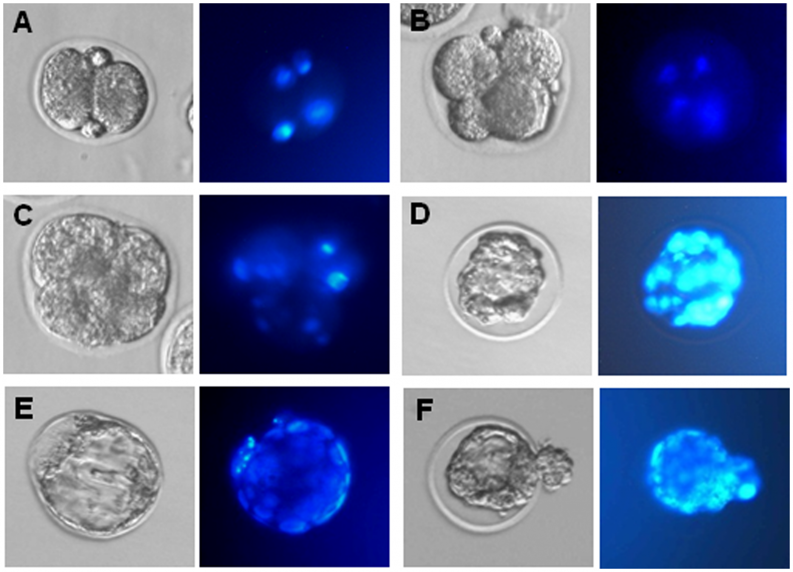
(A–E) Example of 2-cell embryo, 4-cell embryo, morula and blastocyst developed from oocytes generated in vitro in the presence of ActA. Left, phase contrast microscopy observations; right, Hoechst staining of the cell nuclei.
File history
Yi efo/eka'e gwa ebo wo le nyangagi wuncin ye kamina wunga tinya nan
| Gwalagizhi | Nyangagi | Dimensions | User | Comment | |
|---|---|---|---|---|---|
| current | 22:21, 7 August 2012 | | 3,439 × 2,481 (4.19 MB) | Z3330986 (talk | contribs) | (A–E) Example of 2-cell embryo, 4-cell embryo, morula and blastocyst developed from oocytes generated in vitro in the presence of ActA. Left, phase contrast microscopy observations; right, Hoechst staining of the cell nuclei. |
You cannot overwrite this file.
File usage
The following page uses this file: