File:HeartILP draft hearttubesegments.jpg
Original file (1,082 × 771 pixels, file size: 63 KB, MIME type: image/jpeg)
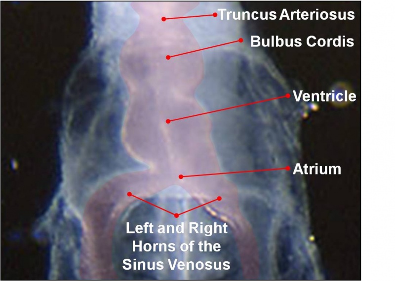
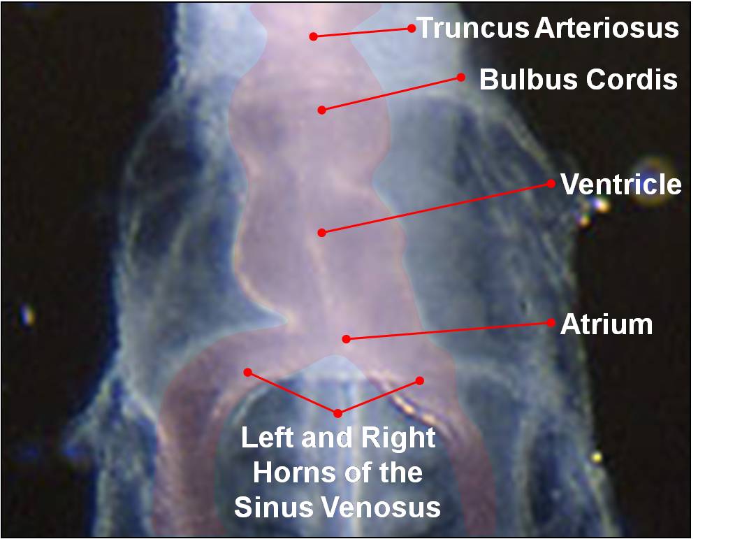
As the tubular heart grows it develops dilations and constrictions which form the truncus arteriosus, bulbus cordis, primitive ventricle, primitive atrium and sinus venosus.
Image Source: Scanning electron micrographs of the Carnegie stages of the early human embryos are reproduced with the permission of Prof Kathy Sulik, from embryos collected by Dr. Vekemans and Tania Attié-Bitach. Images are for educational purposes only and cannot be reproduced electronically or in writing without permission.
File history
Yi efo/eka'e gwa ebo wo le nyangagi wuncin ye kamina wunga tinya nan
| Gwalagizhi | Nyangagi | Dimensions | User | Comment | |
|---|---|---|---|---|---|
| current | 11:15, 22 September 2009 | | 1,082 × 771 (63 KB) | Z3212774 (talk | contribs) | As the tubular heart grows it develops dilations and constrictions which form the truncus arteriosus, bulbus cordis, primitive ventricle, primitive atrium and sinus venosus. {{Template:SEM}} category:HeartILP |
You cannot overwrite this file.
File usage
The following file is a duplicate of this file (more details):
The following page uses this file: