File:Normal control muscle (a) vs. Duchennes muscular dystrophy muscle (b).jpg
_vs._Duchennes_muscular_dystrophy_muscle_(b).jpg)
_vs._Duchennes_muscular_dystrophy_muscle_(b).jpg&diff=cur&oldid=71517)
_vs._Duchennes_muscular_dystrophy_muscle_(b).jpg&direction=next&oldid=71517)
_vs._Duchennes_muscular_dystrophy_muscle_(b).jpg&diff=next&oldid=71517)
Normal_control_muscle_(a)_vs._Duchennes_muscular_dystrophy_muscle_(b).jpg (423 × 550 pixels, file size: 131 KB, MIME type: image/jpeg)
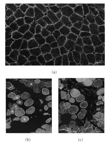
Immunofluorescence with anti-AQP4 antibody of normal control muscle (a) and Duchenne muscular dystrophy muscle (DMD) (b), and that with anti-spectrin antibody of serial muscle section of DMD (c). Positive immunoreactivity with anti-AQP4 antibody is seen in apparently all myofibers of normal control muscles (a); while it is noted in mosaic pattern in DMD muscle (b). DMD muscle contains less numerous myofibers with positive immunoreactivity of anti-AQP4 antibody (b) than myofibers with that of anti-spectrin antibody (c). Scattered anti-AQP4 immunonegative fibers (asterisks in (b)) are noted in DMD muscle. Scale bar in (a)–(c) = 50μm.
Normal control muscle (a) vs. Duchennes muscular dystrophy muscle (b).jpg
Copyright notice: This is an open access article distributed under the Creative Commons Attribution License, which permits unrestricted use, distribution, and reproduction in any medium, provided the original work is properly cited.
File history
Yi efo/eka'e gwa ebo wo le nyangagi wuncin ye kamina wunga tinya nan
| Gwalagizhi | Nyangagi | Dimensions | User | Comment | |
|---|---|---|---|---|---|
| current | 10:27, 22 September 2011 | | 423 × 550 (131 KB) | Z3332629 (talk | contribs) | Immunofluorescence with anti-AQP4 antibody of normal control muscle (a) and Duchenne muscular dystrophy muscle (DMD) (b), and that with anti-spectrin antibody of serial muscle section of DMD (c). Positive immunoreactivity with anti-AQP4 antibody is seen i |
You cannot overwrite this file.
File usage
The following 2 pages use this file:
_vs._Duchennes_muscular_dystrophy_muscle_(b).jpg&oldid=71517)