File:Spinal problems DMD.jpg
Spinal_problems_DMD.jpg (568 × 376 pixels, file size: 127 KB, MIME type: image/jpeg)
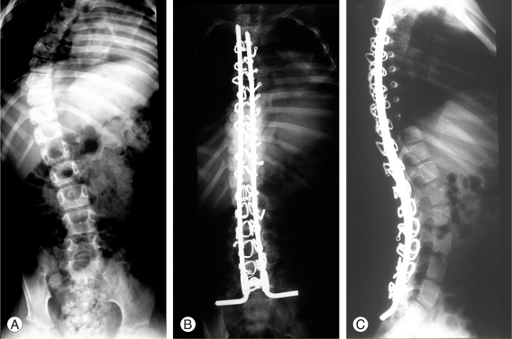
A 11-year-old boy, wheelchair bound for 8 months with Duchenne muscular dystrophy. (A) Preoperative antero-posterior radiograph right sided showing 60° curve. (B, C) Two year post-operative antero-posterior and lateral radiographs showing sublaminar wiring instrumentation with Luque rods and distal fixation to pelvis with L-rod configuration.[1]
Reference
- ↑ <pubmed>21386945</pubmed>
This is an Open Access article distributed under the terms of the Creative Commons Attribution Non-Commercial License (http://creativecommons.org/licenses/by-nc/3.0) which permits unrestricted non-commercial use, distribution, and reproduction in any medium, provided the original work is properly cited.
File history
Yi efo/eka'e gwa ebo wo le nyangagi wuncin ye kamina wunga tinya nan
| Gwalagizhi | Nyangagi | Dimensions | User | Comment | |
|---|---|---|---|---|---|
| current | 22:18, 16 September 2011 |  | 568 × 376 (127 KB) | Z3332824 (talk | contribs) | A 11-year-old boy, wheelchair bound for 8 months with Duchenne muscular dystrophy. (A) Preoperative antero-posterior radiograph right sided showing 60° curve. (B, C) Two year post-operative antero-posterior and lateral radiographs showing sublaminar wiri |
You cannot overwrite this file.
File usage
The following file is a duplicate of this file (more details):
The following 2 pages use this file:
