File:ZUltrasound Image of Fetal Aorta.jpg
ZUltrasound_Image_of_Fetal_Aorta.jpg (640 × 480 pixels, file size: 47 KB, MIME type: image/jpeg)
What am I looking at?
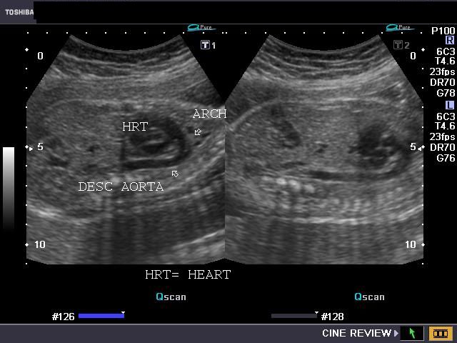
This is a 2D ultrasound image of a fetal chest in sagittal section. Marked with arrows on the image are the fetal heart, aortic arch and descending aorta.
Image Copyright Information
Image obtained at: http://www.ultrasound-images.com/fetal-chest.htm
Author (content provider): Dr. Joe Antony
This image is not classed as a public domain image, but has been reproduced here with the kind permission of Dr. Joe Antony, who controls the website Ultrasound Images, "a free gallery of high-resolution, ultrasound, color Doppler and 3D images". Correspondence attesting to this fact will be cheerfully provided upon request.
File history
Yi efo/eka'e gwa ebo wo le nyangagi wuncin ye kamina wunga tinya nan
| Gwalagizhi | Nyangagi | Dimensions | User | Comment | |
|---|---|---|---|---|---|
| current | 20:10, 10 September 2010 | | 640 × 480 (47 KB) | Z3252833 (talk | contribs) | ===What am I looking at?=== This is a 2D ultrasound image of a fetal chest in sagittal section. Marked with arrows on the image are the fetal heart, aortic arch and descending aorta. ===Image Copyright Information=== Image obtained at: http://www.ul |
You cannot overwrite this file.
File usage
The following page uses this file:


