File:Embryonic dorsal root ganglia in mouse.jpg
From Embryology
No higher resolution available.
Embryonic_dorsal_root_ganglia_in_mouse.jpg (765 × 599 pixels, file size: 84 KB, MIME type: image/jpeg)
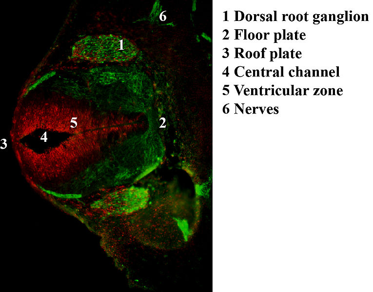
Antibody stain against Neurofilament (green) and Ki 67 (red) in a Mouse embryo at day 12.5 after fertilization. Shown is the dorsal root ganglion (green ellipsoid regions where cells express neurofilament) and the ventricular zone (red region where cells proliferate) as well as the neural tube with roof and floor plate.
File history
Yi efo/eka'e gwa ebo wo le nyangagi wuncin ye kamina wunga tinya nan
| Gwalagizhi | Nyangagi | Dimensions | User | Comment | |
|---|---|---|---|---|---|
| current | 19:50, 16 October 2018 | | 765 × 599 (84 KB) | Z5229431 (talk | contribs) | Antibody stain against Neurofilament (green) and Ki 67 (red) in a Mouse embryo at day 12.5 after fertilization. Shown is the dorsal root ganglion (green ellipsoid regions where cells express neurofilament) and the ventricular zone (red region where cel... |
You cannot overwrite this file.
File usage
The following 2 pages use this file:


