File:Structure of DRG in Sox10-deficient mice.jpg
From Embryology

Size of this preview: 282 × 598 pixels. Other resolution: 969 × 2,055 pixels.
Original file (969 × 2,055 pixels, file size: 441 KB, MIME type: image/jpeg)
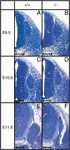
Histology of DRG in Sox10-deficient mice. Transverse sections, 6 μm through the thoracic neural tube and DRG from 9.5 to 11.5 dpc embryos were stained with toluduine blue. Wildtype (+/+ in A, C, E) and homozygous Sox10-deficient embryos (−/− in B, D, F). The arrowhead (F) marks the connection of DRG with dorsal neural tube. Bars represent 100 μm.
File history
Click on a date/time to view the file as it appeared at that time.
| Date/Time | Thumbnail | Dimensions | User | Comment | |
|---|---|---|---|---|---|
| current | 14:49, 15 October 2018 | | 969 × 2,055 (441 KB) | Z5229431 (talk | contribs) | Histology of DRG in Sox10-deficient mice. Transverse sections, 6 μm through the thoracic neural tube and DRG from 9.5 to 11.5 dpc embryos were stained with toluduine blue. Wildtype (+/+ in A, C, E) and homozygous Sox10-deficient embryos (−/− in B,... |
You cannot overwrite this file.
File usage
There are no pages that use this file.