File:3D Organoids.jpg
From Embryology
Size of this preview: 800 × 354 pixels. Other resolution: 1,600 × 708 pixels.
Original file (1,600 × 708 pixels, file size: 1.11 MB, MIME type: image/jpeg)
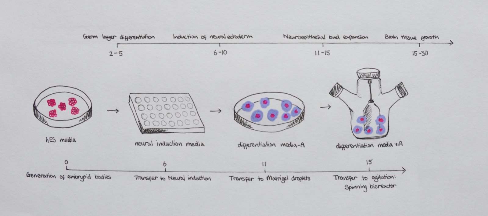
Timeline and method of cerebral organoid development
Description
Timeline of the protocol involved in development of 3D cerebral organoids from human pluripotent stem cells, first aggregating to for embryoid bodies.
Copyright
"Beginning six months after publication, I (z5178570) grant the public the non-exclusive right to copy, distribute, or display the Work under a Creative Commons Attribution-Noncommercial-Share Alike 3.0 Unported license, as described at http://creativecommons.org/licenses/by-nc-sa/3.0/ and http://creativecommons.org/licenses/by-nc-sa/3.0/legalcode."
Reference
Hand drawn student image, based upon schematic from: [1]
- ↑ <pubmed>25188634</pubmed>
File history
Click on a date/time to view the file as it appeared at that time.
| Date/Time | Thumbnail | Dimensions | User | Comment | |
|---|---|---|---|---|---|
| current | 19:38, 23 October 2017 | | 1,600 × 708 (1.11 MB) | Z5178570 (talk | contribs) | Hand drawn student image, based upon schematic from: Madeline A Lancaster, Juergen A Knoblich Generation of cerebral organoids from human pluripotent stem cells. Nat Protoc: 2014, 9(10);2329-40 |
You cannot overwrite this file.
File usage
The following 2 pages use this file: