File:Cortical plate development.jpg
Original file (1,600 × 2,648 pixels, file size: 4.02 MB, MIME type: image/jpeg)
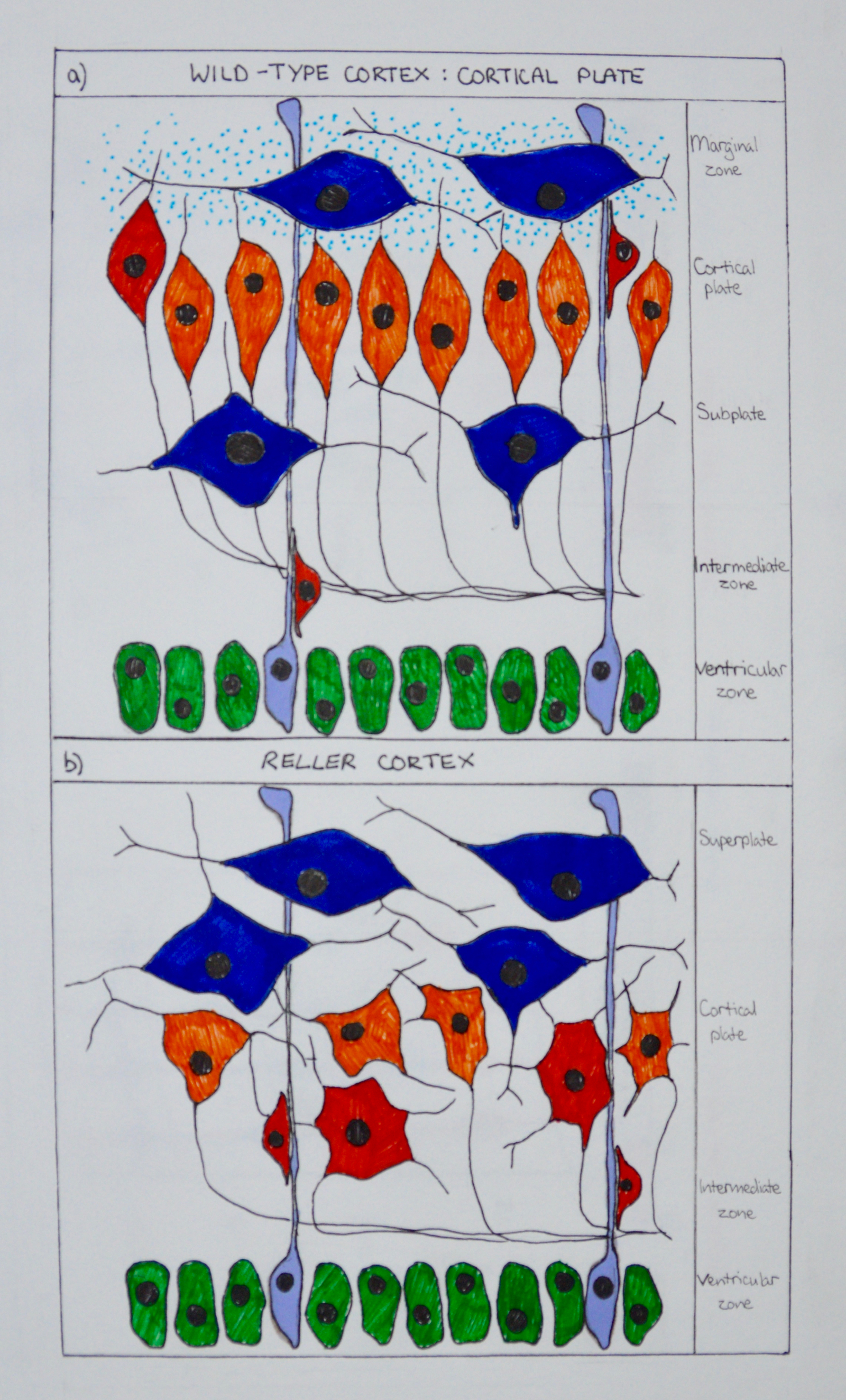
Preplate stage of cerebral cortex development. Preplate neurons are dark blue; undifferentiated ventricular zone cells are green; radial glia are purple; and Reelin, produced by Cajal-Retzius cells, is represented by light blue dots. (a) Separation of the preplate into subplate and marginal zone by the developing cortical plate in wild-type mice. Future layer VI neurons are orange; future layer V neurons are red. (b) In reeler mutant mice, the developing cortical plate fails to separate the preplate into two layers. Neurons that should become layer V (red) do not by pass the neurons that should become layer VI (yellow). In the reeler cortex, the dendritic processes are arranged in a haphazard manner, whereas the axons are mostly normal.
File history
Yi efo/eka'e gwa ebo wo le nyangagi wuncin ye kamina wunga tinya nan
| Gwalagizhi | Nyangagi | Dimensions | User | Comment | |
|---|---|---|---|---|---|
| current | 14:18, 22 October 2017 | | 1,600 × 2,648 (4.02 MB) | Z5178570 (talk | contribs) | Preplate stage of cerebral cortex development. Preplate neurons are dark blue; undifferentiated ventricular zone cells are green; radial glia are purple; and Reelin, produced by Cajal-Retzius cells, is represented by light blue dots. (a) Separation of... |
You cannot overwrite this file.
File usage
The following 2 pages use this file: