File:Amphioxus development.gif
Original file (778 × 884 pixels, file size: 284 KB, MIME type: image/gif)
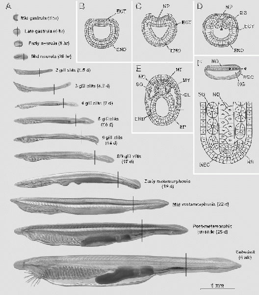
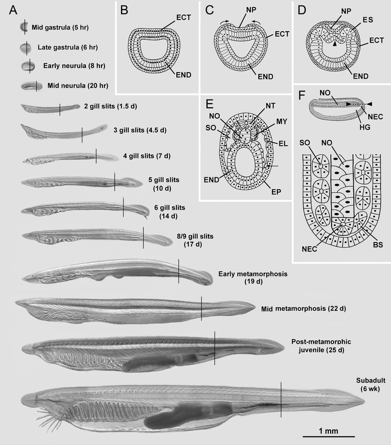
Overview of amphioxus development and the stages examined in this study. (A) Differential interference contrast micrographs of living specimens in side view with anterior to the left (with times after fertilization); the present study was limited to studying the body region indicated by the vertical line on each specimen; the 1-mm scale line applies to all images. (B-E) Schematic drawings of transverse sections of embryos and early larvae. (B) Late gastrula. (C) Gastrula-to-neurula transition (arrows indicate epidermis overgrowing neural plate during the first phase of neurulation). (D) Early neurula (arrowhead indicates initial stage of evagination of the notochord mid-dorsally from the endoderm). (E) Mid neurula (arrow indicates the ventral somitocoel extension that later pinches off to give rise to the perivisceral coelom). (F) Formation of more posterior somites occurs through segmentation from the neurenteric canal. Drawings B-E are reproduced from Hatschek [14,27], respectively. Abbreviations: BS, budding somite; ECT, ectoderm; EL, external cell layer; END, endoderm; EP, epidermis; ES, epithelial somite; HG, hindgut; MY, myotome; NEC, neurenteric canal; NO, notochord; NP, neural plate; NT, neural tube.
File history
Yi efo/eka'e gwa ebo wo le nyangagi wuncin ye kamina wunga tinya nan
| Gwalagizhi | Nyangagi | Dimensions | User | Comment | |
|---|---|---|---|---|---|
| current | 10:50, 24 October 2016 | | 778 × 884 (284 KB) | Z3516832 (talk | contribs) | Overview of amphioxus development and the stages examined in this study. (A) Differential interference contrast micrographs of living specimens in side view with anterior to the left (with times after fertilization); the present study was limited to st... |
You cannot overwrite this file.
File usage
The following 2 pages use this file: