File:Hypothyroidism.jpg

Original file (1,492 × 1,634 pixels, file size: 565 KB, MIME type: image/jpeg)
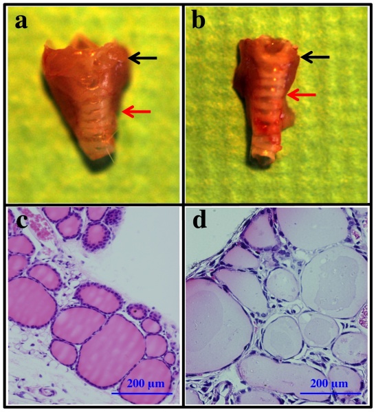
This image compares a normal thyroid gland (a) anatomy with a hypothyroidism thyroid gland (b). The histology images reveal in the abnormal thyroid gland, there is less thyroid hormone produced due to irregular epithelium on the surface of the follicles. The researchers in this report found that once thyroid stimulating hormone was added to the hypothyroid follicles, the epithelium became more columnar and the lumens became smaller, similar to healthy thyroid follicles. Cite error: Invalid <ref> tag; invalid names, e.g. too many
http://www.plosone.org/article/info%3Adoi%2F10.1371%2Fjournal.pone.0042358
© Endo, Kobayashi. This is an open-access article distributed under the terms of the Creative Commons Attribution License, which permits unrestricted use, distribution, and reproduction in any medium, provided the original author and source are credited.
File history
Click on a date/time to view the file as it appeared at that time.
| Date/Time | Thumbnail | Dimensions | User | Comment | |
|---|---|---|---|---|---|
| current | 09:18, 24 October 2014 | | 1,492 × 1,634 (565 KB) | Z3414648 (talk | contribs) | This image compares a normal thyroid gland (a) anatomy with a hypothyroidism thyroid gland (b). The histology images reveal in the abnormal thyroid gland, there is less thyroid hormone produced due to irregular epithelium on the surface of the follicle... |
You cannot overwrite this file.
File usage
The following 2 pages use this file: