File:Anatomical diagram of testes.jpg
From Embryology
Size of this preview: 465 × 599 pixels. Other resolution: 647 × 834 pixels.
Original file (647 × 834 pixels, file size: 237 KB, MIME type: image/jpeg)
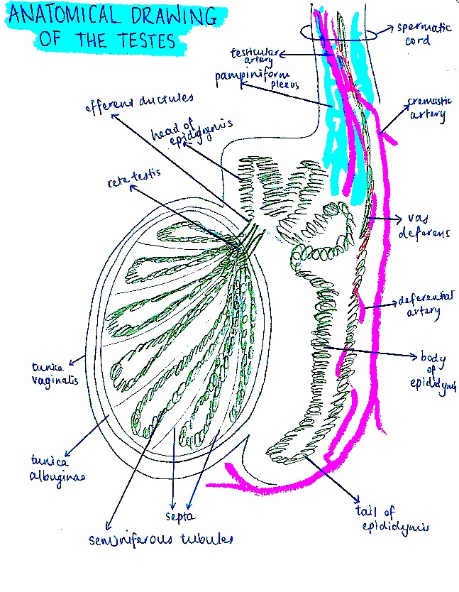
Student drawn image of the testes, epididymis, ductus deferens and spermatic cord.
File history
Yi efo/eka'e gwa ebo wo le nyangagi wuncin ye kamina wunga tinya nan
| Gwalagizhi | Nyangagi | Dimensions | User | Comment | |
|---|---|---|---|---|---|
| current | 21:29, 20 October 2014 | | 647 × 834 (237 KB) | Z3417753 (talk | contribs) | Reverted to version as of 10:27, 20 October 2014 |
| 21:28, 20 October 2014 | 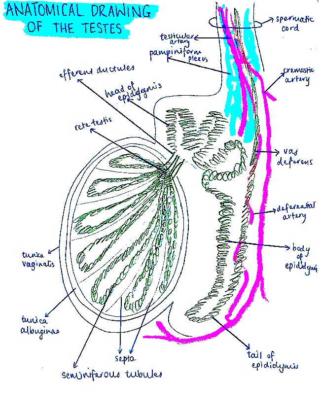 | 647 × 834 (237 KB) | Z3417753 (talk | contribs) | Reverted to version as of 10:08, 20 October 2014 | |
| 21:27, 20 October 2014 | 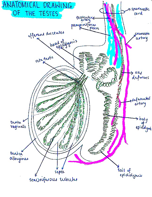 | 647 × 834 (237 KB) | Z3417753 (talk | contribs) | Reverted to version as of 10:08, 20 October 2014 | |
| 21:08, 20 October 2014 | 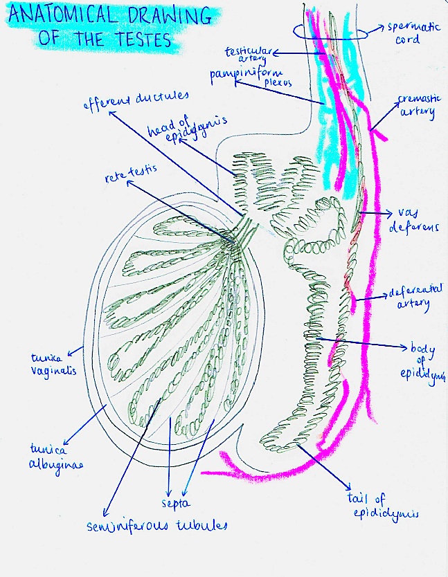 | 647 × 834 (235 KB) | Z3417753 (talk | contribs) | Reverted to version as of 10:05, 20 October 2014 | |
| 21:08, 20 October 2014 | 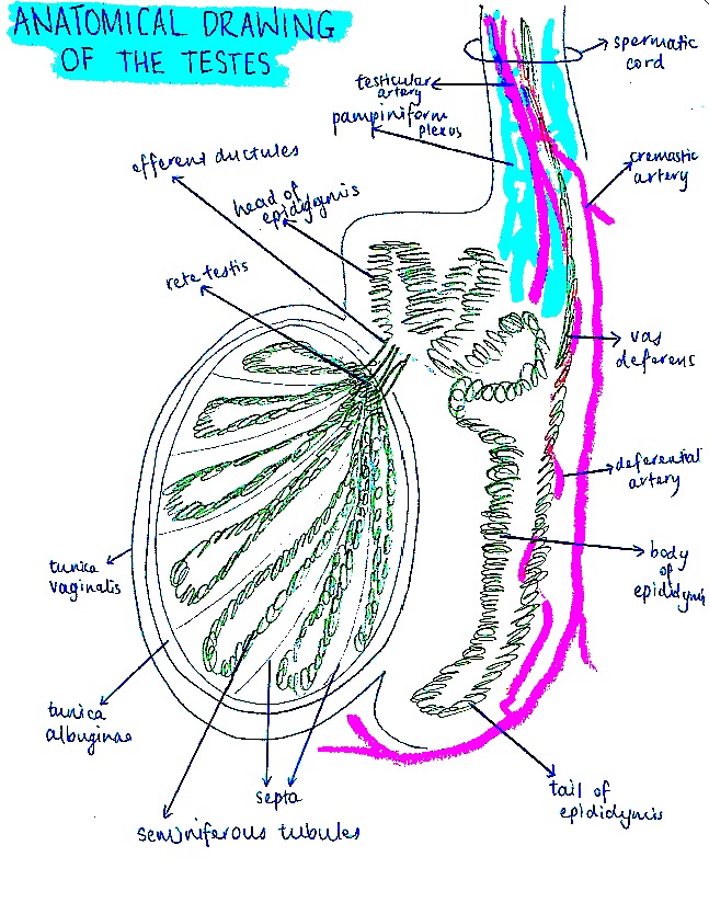 | 647 × 834 (237 KB) | Z3417753 (talk | contribs) | ||
| 21:05, 20 October 2014 | 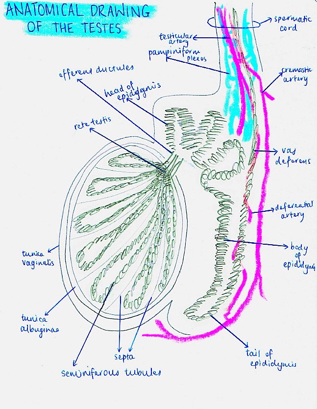 | 647 × 834 (235 KB) | Z3417753 (talk | contribs) | ||
| 20:24, 20 October 2014 |  | 647 × 834 (472 KB) | Z3417753 (talk | contribs) | Student drawn image of the testes, epididymis, ductus deferens and spermatic cord. |
You cannot overwrite this file.
File usage
The following 2 pages use this file:


