File:Secondary Oocyte during in-vitro fertilisation.jpeg
From Embryology
No higher resolution available.
Secondary_Oocyte_during_in-vitro_fertilisation.jpeg (800 × 558 pixels, file size: 48 KB, MIME type: image/jpeg)
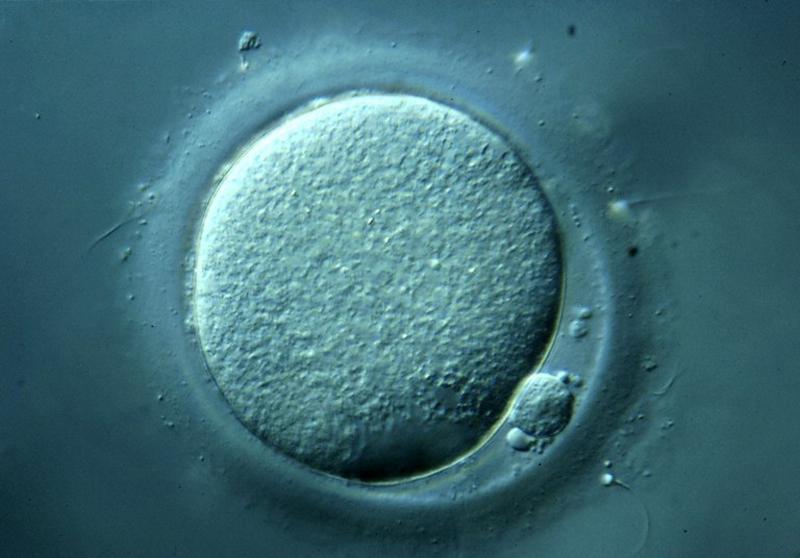
A human secondary oocyte during in-vitro fertilisation viewed with a light microscope. The small cell at the bottom right is a polar body. Spice Walker, Wellcome Images.
z3418698
File history
Yi efo/eka'e gwa ebo wo le nyangagi wuncin ye kamina wunga tinya nan
| Gwalagizhi | Nyangagi | Dimensions | User | Comment | |
|---|---|---|---|---|---|
| current | 21:48, 20 August 2014 | | 800 × 558 (48 KB) | Z3418698 (talk | contribs) | A human secondary oocyte during in-vitro fertilisation viewed with a light microscope. The small cell at the bottom right is a polar body. Spice Walker, Wellcome Images. z3418698 |
You cannot overwrite this file.
File usage
There are no pages that use this file.


