File:E18.5 developing kidney expressing Pygo1 and Pygo2.jpg
From Embryology

Size of this preview: 800 × 525 pixels. Other resolution: 1,200 × 787 pixels.
Original file (1,200 × 787 pixels, file size: 260 KB, MIME type: image/jpeg)
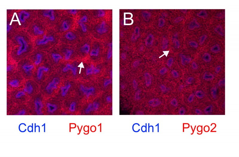
Expression patterns of Pygo1 and Pygo2 proteins in the cortex of E18.5 kidney was determined using immunofluorescence. The location of both Pygo1 and Pygo2 were in the nucleus with the colour red. Both genes are expressed widely where in all the components of the developing kidney, a signal is detected. However their were high levels of stromal cell compartment(arrows). Original magnification x200
Student number: 3414515
File history
Yi efo/eka'e gwa ebo wo le nyangagi wuncin ye kamina wunga tinya nan
| Gwalagizhi | Nyangagi | Dimensions | User | Comment | |
|---|---|---|---|---|---|
| current | 09:34, 20 August 2014 |  | 1,200 × 787 (260 KB) | Z3414515 (talk | contribs) | Expression patterns of Pygo1 and Pygo2 proteins in the cortex of E18.5 kidney was determined using immunofluorescence. The location of both Pygo1 and Pygo2 were in the nucleus with the colour red. Both genes are expressed widely where in all the compon... |
You cannot overwrite this file.
File usage
The following page uses this file: