File:Fusion of two pairs of blastomeres inside 4-cell embryos.png
From Embryology

Size of this preview: 798 × 600 pixels. Other resolution: 1,247 × 937 pixels.
Original file (1,247 × 937 pixels, file size: 1.39 MB, MIME type: image/png)
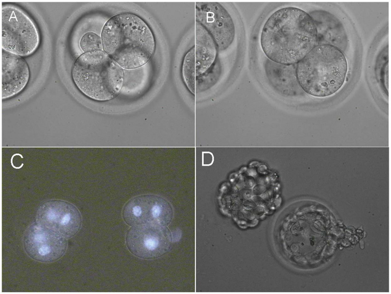
(A) Fusion of first pair of blastomeres. (B) Fusion of second pair of blastomeres. (C) Resulting 2-cell embryos. Hoechst 33342 staining confirms the presence of two nuclei in each blastomere. (D) In vitro development of 4-cell embryos with two fused pairs of blastomeres. Hatching and hatched blastocyst.
<pubmed>23227157</pubmed>
- Note - This image was originally uploaded as part of an undergraduate science student project and may contain inaccuracies in either description or acknowledgements. Students have been advised in writing concerning the reuse of content and may accidentally have misunderstood the original terms of use. If image reuse on this non-commercial educational site infringes your existing copyright, please contact the site editor for immediate removal.
File history
Click on a date/time to view the file as it appeared at that time.
| Date/Time | Thumbnail | Dimensions | User | Comment | |
|---|---|---|---|---|---|
| current | 14:27, 19 August 2014 | | 1,247 × 937 (1.39 MB) | Z3417753 (talk | contribs) | (A) Fusion of first pair of blastomeres. (B) Fusion of second pair of blastomeres. (C) Resulting 2-cell embryos. Hoechst 33342 staining confirms the presence of two nuclei in each blastomere. (D) In vitro development of 4-cell embryos with two fused pa... |
You cannot overwrite this file.
File usage
The following page uses this file: