File:Week6.jpg
From Embryology
Size of this preview: 545 × 599 pixels. Other resolution: 621 × 683 pixels.
Original file (621 × 683 pixels, file size: 89 KB, MIME type: image/jpeg)
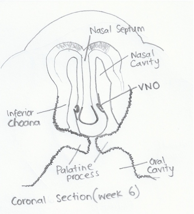
A diagram of the coronal section of an embryo at week 6 of development, indicating the formation of the vomeronasal organ, choana and palatine processes. VNO: Vomeronasal Organ
Image is self drawn by Student based on histology provided by: <pubmed>15454774</pubmed>
- Note - This image was originally uploaded as part of an undergraduate science student project and may contain inaccuracies in either description or acknowledgements. Students have been advised in writing concerning the reuse of content and may accidentally have misunderstood the original terms of use. If image reuse on this non-commercial educational site infringes your existing copyright, please contact the site editor for immediate removal.
File history
Yi efo/eka'e gwa ebo wo le nyangagi wuncin ye kamina wunga tinya nan
| Gwalagizhi | Nyangagi | Dimensions | User | Comment | |
|---|---|---|---|---|---|
| current | 02:43, 3 October 2012 | | 621 × 683 (89 KB) | Z3331264 (talk | contribs) |
You cannot overwrite this file.
File usage
The following 2 pages use this file: