File:MRI Goldenhar Syndrome.jpg
From Embryology
No higher resolution available.
MRI_Goldenhar_Syndrome.jpg (256 × 256 pixels, file size: 8 KB, MIME type: image/jpeg)
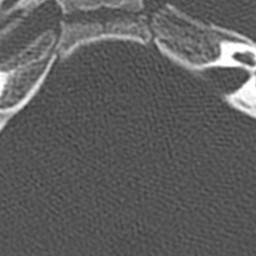
CT scans of inner ear cavities and adjacent sinuses performed at Texas Children’s Hospital. A, Case Presentation 1 showing diminished left middle ear cavity and mastoid sinus (yellow arrow)
Reference: <pubmed>PMC3441207</pubmed>
Copyright: ©2012 This is an Open Access article distributed under the terms of the Creative Commons Attribution License, which permits unrestricted use, distribution, and reproduction in any medium, provided the original work is properly cited.
File history
Yi efo/eka'e gwa ebo wo le nyangagi wuncin ye kamina wunga tinya nan
| Gwalagizhi | Nyangagi | Dimensions | User | Comment | |
|---|---|---|---|---|---|
| current | 16:41, 30 September 2012 | | 256 × 256 (8 KB) | Z3292017 (talk | contribs) | CT scans of inner ear cavities and adjacent sinuses performed at Texas Children’s Hospital. A, Case Presentation 1 showing diminished left middle ear cavity and mastoid sinus (yellow arrow) Reference: <pubmed>PMC3441207</pubmed> Copyright: ©2012 This |
You cannot overwrite this file.
File usage
There are no pages that use this file.