Unused files
From Embryology
The following files exist but are not embedded in any page. Please note that other web sites may link to a file with a direct URL, and so may still be listed here despite being in active use.
Showing below up to 50 results in range #951 to #1,000.
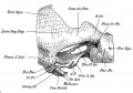 Watson1915 Fig02.jpg 1,421 × 1,000; 245 KB
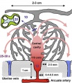 Placenta lobule blood flow cartoon.jpg 692 × 800; 134 KB
 Ziegler model legend 33.jpg 800 × 800; 74 KB
 Ziegler model legend 35.jpg 800 × 800; 60 KB
 Ziegler model legend 37.jpg 800 × 800; 51 KB
 Ziegler model legend 38.jpg 800 × 800; 53 KB
 Ziegler model legend 40.jpg 800 × 800; 0 bytes
 Ziegler model legend 41.jpg 800 × 800; 64 KB
 Ziegler model legend 45.jpg 800 × 799; 0 bytes
 Ziegler model legend 48.jpg 800 × 803; 74 KB
 Axolotl limb joints 01.jpg 1,200 × 588; 408 KB
 Embryology website 1996.jpg 764 × 600; 128 KB
 Wilson Hill Martin.jpg 800 × 548; 0 bytes
 B100658-01.jpg 1,388 × 1,040; 677 KB
 Embryo 1.6mm-1.gif 299 × 400; 299 KB
 Embryo 3.1mm-1.gif 299 × 400; 238 KB
 Embryo 10mm surface-1.gif 299 × 400; 393 KB
 2013 Wiki Stats.xls ; 28 KB
 Chicken primitive streak cell fate 02.jpg 468 × 661; 38 KB
 Hochstadter plate 16.jpg 1,507 × 2,000; 640 KB
 IBook sales 2013.jpg 800 × 367; 28 KB
 Chicken limb gene expression 01.jpg 1,200 × 758; 85 KB
 Stage16 em11.gif 800 × 456; 336 KB
 SHPracManual 2014 Health Safety.pdf ; 120 KB
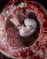 Stage14 bf19.jpg 1,200 × 1,500; 287 KB
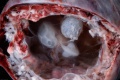 Stage14 bf22.jpg 1,500 × 1,001; 214 KB
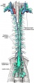 Gray599-1.jpg 351 × 850; 98 KB
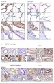 Human developing lung protein 01.jpg 657 × 1,000; 241 KB
 Human developing lung protein 02.jpg 800 × 302; 82 KB
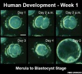 Human blastocyst day 1-5.jpg 500 × 450; 46 KB
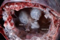 Stage14 bf25.jpg 2,000 × 1,334; 337 KB
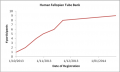 HFTB graph 1.png 482 × 289; 24 KB
 HFTB graph 3.png 609 × 360; 21 KB
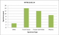 HFTB graph 2.png 609 × 365; 12 KB
 Stage5 bf13.jpg 800 × 537; 142 KB
 Placenta percreta 02.jpg 1,000 × 405; 105 KB
 HillH202 Stage 17 600.gif 600 × 600; 346 KB
 HillH202 Stage 17 bf03.jpg 800 × 800; 86 KB
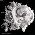 HillH202 Stage 17 bf04.jpg 1,000 × 1,000; 138 KB
 Parotid gland histology 02.jpg 479 × 600; 95 KB
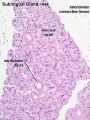 Sublingual gland histology 03.jpg 400 × 533; 73 KB
 Submandibular gland histology 01.jpg 1,280 × 1,024; 390 KB
 Submandibular gland histology 02.jpg 1,280 × 1,024; 400 KB
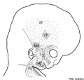 Embryonic neck muscle cartoon.jpg 600 × 570; 45 KB
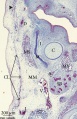 Human embryo neck 01.jpg 534 × 827; 186 KB
 Human fetal neck 01.jpg 549 × 827; 186 KB
 Search embryology 2014-04-02.jpg 1,000 × 557; 132 KB
 Spermatozoa mitochondria PMID23878233.gif 495 × 495; 974 KB
 Thyng1914 plate01.jpg 1,147 × 1,500; 370 KB
 George Streeter.jpg 318 × 466; 21 KB


