Unused files
From Embryology
The following files exist but are not embedded in any page. Please note that other web sites may link to a file with a direct URL, and so may still be listed here despite being in active use.
Showing below up to 50 results in range #651 to #700.
 Human embryo skin 8-9 week EGA desmosomes.jpg 800 × 198; 40 KB
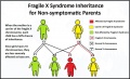 Fragile X Inheritance..jpg 1,074 × 654; 105 KB
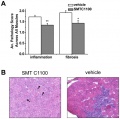 Tinsley et al. utrophin results.jpg 452 × 449; 114 KB
 Experiment groups for utrophin upregulation.JPG 932 × 128; 20 KB
 2011 Lecture - Neural Development.pdf ; 3.08 MB
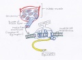 Dystrophin in the muscle fibre membrane.jpg 1,840 × 1,355; 415 KB
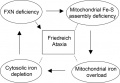 Role of FXN Gene in Friedriech Ataxia.jpg 514 × 356; 51 KB
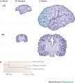 Species brain sizes.jpg 600 × 664; 87 KB
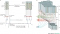 Model of cortical neurogenesis.jpg 1,000 × 565; 99 KB
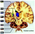 Regions of the brain.jpg 1,280 × 1,239; 348 KB
- ANAT2341 2011 Heart Dunwoodie.pdf ; 1.3 MB
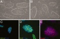 Morula cell lines express ES markers.jpg 1,280 × 831; 205 KB
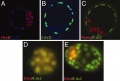 Early lineage markers in morulae and blastocysts.jpg 1,280 × 868; 114 KB
 Embryology qrurl.jpg 328 × 328; 3 KB
 Site map qrurl.jpg 392 × 392; 5 KB
 Medicine qrurl.jpg 392 × 392; 5 KB
 Science qrurl.jpg 392 × 392; 5 KB
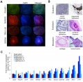 Cord blood induced stem cells 01.jpg 800 × 787; 157 KB
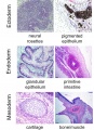 Cord blood induced stem cells 02.jpg 700 × 966; 189 KB
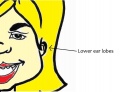 Lowered Ear Lobes.jpg 1,078 × 828; 72 KB
 Kollmann499.jpg 550 × 367; 40 KB
 Kollmann500.jpg 548 × 344; 38 KB
 Kollmann497-498.jpg 762 × 277; 45 KB
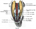 Kollmann565.jpg 703 × 571; 79 KB
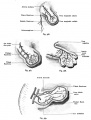 Kollmann566-569.jpg 815 × 1,074; 112 KB
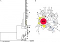 Dog mitochondrial DNA variations.jpg 1,000 × 703; 76 KB
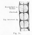 Kollmann051.jpg 327 × 354; 16 KB
 Microtiter plate - 96 wells.jpg 530 × 351; 67 KB
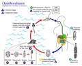 Opisthorchiasis.jpg 739 × 598; 84 KB
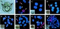 Human preimplantation embryos 01.jpg 1,280 × 629; 261 KB
 Mouse interdigit apoptosis 02.jpg 764 × 764; 61 KB
 Bones.pptx ; 64 KB
 Rudolf Virchow.jpg 480 × 656; 170 KB
- 2011 Wiki Stats.xls ; 21 KB
 Gray0001.jpg 2,048 × 1,052; 185 KB
 Gray0002.jpg 963 × 1,536; 111 KB
 Gray0004.jpg 2,048 × 1,205; 199 KB
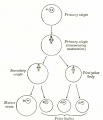 Gray0005.jpg 1,314 × 1,536; 66 KB
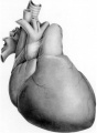 Bedford01.jpg 734 × 1,000; 82 KB
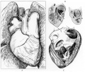 Bedford02.jpg 1,069 × 911; 266 KB
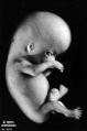 Stage23 bf7.jpg 600 × 906; 23 KB
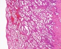 Uterus secretory phase 01.jpg 1,280 × 1,024; 318 KB
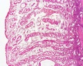 Uterus secretory phase 02.jpg 1,280 × 1,024; 359 KB
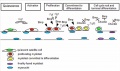 Model BMP satellite cell differentiation.jpg 800 × 473; 75 KB
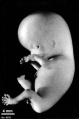 Stage23 bf8.jpg 578 × 872; 43 KB
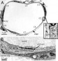 Venule microvessel EM.jpg 600 × 626; 91 KB
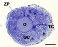 Mouse in vitro follicle 01.jpg 661 × 534; 72 KB
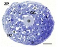 Mouse in vitro follicle 02.jpg 800 × 639; 120 KB
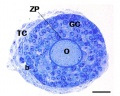 Mouse in vitro follicle 03.jpg 800 × 639; 99 KB


