Search results
From Embryology
File:Z3333865.implantation.png Su R-W, Jia B, Ni H, Lei W, Yue S-L, et al. (2012) '''Junctional Adhesion Molecule 2 Mediates the Inte(920 × 2,546 (2.87 MB)) - 07:28, 8 August 2012
File:Rugh 044.jpg Courtesy, W. R. Duryee, 1950, Ann. N. Y. Acad. Sci., 50, Art. 8.(882 × 1,000 (226 KB)) - 09:13, 12 April 2013
File:Fawcett1975 fig34.jpg From D. W. Fawcett, D. S. Friend, M. Price and R. W. Linck, Proc. 8th Intemat. Congr. Electron Microscopy, Canberra, 1974(1,280 × 1,746 (506 KB)) - 09:58, 20 August 2017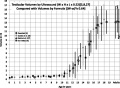
File:Testicular volume graph.jpg ...groups compared with volumes in subjects obtained clinically by formula ((W-ss)3 × 0.64).(554 × 405 (57 KB)) - 13:41, 3 June 2018
File:Wen1928-Fig09.jpg We are indebted to Dr. G. W. Corner for suggesting this type of reconstruction.(755 × 1,000 (91 KB)) - 15:31, 20 April 2016
File:Rugh 069.jpg After W. Vogt, 1929b. From Spemann: "Embryonic Development and Induction," New Have(1,200 × 603 (201 KB)) - 12:34, 12 April 2013
File:Keibel Mall 2 330.jpg ...of the thymus; Thyr., thyreoid; ub. K., ultimobranchial body; Vag., vagus; W., vertebra. X 60.(1,280 × 1,009 (342 KB)) - 13:50, 12 January 2017
File:Bailey041.jpg A, anterior end of the blastoderm; G. W., germ wall; M, yolk masses of the blastocoel; P, posterior margin of blast(1,071 × 283 (103 KB)) - 23:30, 13 February 2011
File:Corner-table01.jpg By George W. Corner. (4 plates, 2 text-figures) 117-146(600 × 335 (36 KB)) - 12:14, 24 February 2011
File:Corner-plate03.jpg By George W. Corner. (4 plates, 2 text-figures) 117-146(744 × 960 (150 KB)) - 12:18, 24 February 2011
File:Keibel1907 titlepage.jpg ...''Farsius spectrum'') and slow loris (''Nycticebus tardigradus'') by A. A. W. Hubrecht and Franz Keibel(685 × 1,000 (76 KB)) - 18:17, 24 January 2014
File:Bailey027.jpg D, from the vegetal pole, c, Gray crescent; w, nonpigmented vegetal pole.(553 × 481 (87 KB)) - 18:45, 16 March 2011
File:Hertig1946b fig12a.jpg ...lining epithelium. (Fig. 5 from Cullen’s “The Umbilicus and Its Diseases,” W. B. Saunders Company.)(800 × 858 (169 KB)) - 08:44, 8 August 2017
File:Corner-plate02.jpg By George W. Corner. (4 plates, 2 text-figures) 117-146(729 × 986 (152 KB)) - 12:19, 24 February 2011
File:Corner-plate04.jpg By George W. Corner. (4 plates, 2 text-figures) 117-146(719 × 974 (193 KB)) - 12:18, 24 February 2011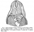
File:Mall1891 Fig01.jpg * iP. W. - foramen of Winslow(623 × 576 (99 KB)) - 16:28, 18 August 2015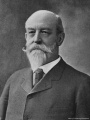
File:Henry Pickering Bowditch.jpg ...ng them F. W. Ellis, G. S. Hall, W.P. Lombard, C. S. Minot, I. Ott, and J. W. Warren, had worked with him in his laboratory. His research touched on a w(750 × 1,000 (129 KB)) - 00:54, 18 July 2015
File:Keibel Mall 001.jpg (From W. Nasel' Arch. f. mikr. Ajiat., vol. mi, 1888, PI. XX, Tit. 6.)(781 × 733 (138 KB)) - 07:00, 23 August 2012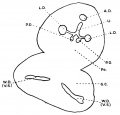
File:Evatt1909 fig05.jpg ...P.D., L.D., anterior posterior, and lateral prostatic ducts respectively; W. D. (V .S.), Wolfilan ducts (vesiculae seminales).(800 × 769 (55 KB)) - 13:33, 17 June 2017
File:Rugh 039.jpg Courtesy, W. R. Duryee, 1950, Ann. N. Y. Acad. ScL, 50, Art. 8.(753 × 800 (63 KB)) - 08:51, 12 April 2013


