Unused files
From Embryology
The following files exist but are not embedded in any page. Please note that other web sites may link to a file with a direct URL, and so may still be listed here despite being in active use.
Showing below up to 50 results in range #501 to #550.
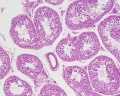 Testis, young H&E reproductive system, male, convoluted seminiferous tubules x10.jpg 1,280 × 1,024; 396 KB
 Mouse follicle in vitro 02.jpg 600 × 701; 146 KB
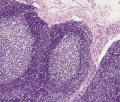
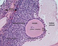
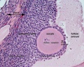
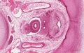
 Mouse antral follicle.jpg 600 × 705; 168 KB
 Mouse adult lymph node 07.mov ; 309 KB
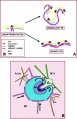
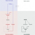
 Model coupling hematopoiesis with osteopoiesis.jpg 486 × 600; 44 KB
 Lymph node histology01.jpg 800 × 680; 282 KB
 Pig lung alveolarization.jpg 600 × 389; 38 KB
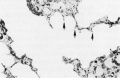
 Cytoplasmic lattices in oocytes and two-cell embryos.jpg 753 × 1,000; 226 KB
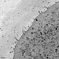
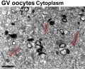 Cytoplasmic lattices in GV oocyte cytoplasm.jpg 1,048 × 846; 291 KB
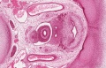
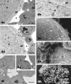 Mouse antral follicle 01.jpg 932 × 1,095; 374 KB
 Model for granulocytic nuclear lobulation.jpg 600 × 930; 85 KB
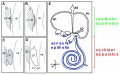
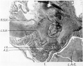
 Sabin1909 fig17.jpg 645 × 545; 80 KB
 Sabin1909 fig13.jpg 640 × 547; 114 KB
 Sabin1909 fig16.jpg 512 × 438; 120 KB
 Human zygote two pronuclei 02.png 433 × 422; 121 KB
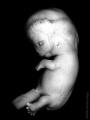
 Canine embryo E21 image002.jpg 600 × 450; 48 KB
 Mouse olfactory development E12-E15.jpg 600 × 1,764; 323 KB
 Bronchial epithelial bridge.jpg 397 × 482; 28 KB
 Stage22 bf2a.jpg 600 × 800; 36 KB
 Stage22 bf2b.jpg 450 × 600; 22 KB
 Stage22 bf2c.jpg 300 × 400; 11 KB
- Intestinal rotation.mov ; 36 KB
 BGDA PracManual 2011 Practical 3.pdf ; 349 KB
 Mouse ovary 01.jpg 602 × 482; 69 KB
 Rat ovary follicle development 01.jpg 1,200 × 1,035; 201 KB
 Human fetal ovary SMAD6 expression.jpg 711 × 535; 167 KB
 Mouse oocyte and zona pellucida EM01b.jpg 600 × 600; 133 KB
 Mouse oocyte and zona pellucida EM01c.jpg 400 × 400; 60 KB
 Mouse inner ear development 01.jpg 600 × 373; 64 KB
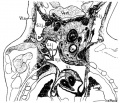
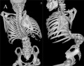
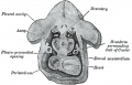 Gray0054.jpg 800 × 513; 71 KB
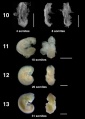 Bat embryo stage 10-13.jpg 547 × 767; 23 KB
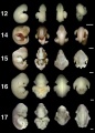 Bat embryo stage 12-17.jpg 548 × 767; 30 KB
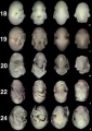 Bat embryo stage 18-24.jpg 518 × 734; 32 KB
 Lens-neural crest signaling 02.jpg 521 × 522; 22 KB
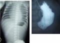 Pyloric atresia 01.jpg 800 × 580; 35 KB
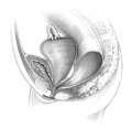 Hydrocolpos.jpg 375 × 361; 23 KB
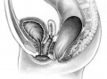 Perineal fistula.jpg 800 × 596; 82 KB
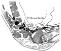
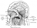 Gray1042.jpg 969 × 745; 140 KB
 Midgut rotation icon.jpg 322 × 241; 17 KB
 Pancreas histology 10he.jpg 300 × 400; 73 KB
 Pancreas histology 40he.jpg 300 × 400; 42 KB
 Spina bifida.jpg 800 × 633; 77 KB
 Liver plasmodium infection cartoon.jpg 1,000 × 450; 78 KB
 Ovary histology 061a.jpg 800 × 640; 200 KB
 Ovary histology 061c.jpg 400 × 320; 56 KB
 Stage 22 image 210a.jpg 1,000 × 644; 310 KB
 Stage 22 image 210b.jpg 800 × 515; 207 KB
 Stage 22 image 210c.jpg 400 × 257; 52 KB
 Platypus right auricle 01.jpg 800 × 640; 124 KB
 Hertwig1892-plate01.jpg 919 × 1,475; 257 KB


