Search results
From Embryology
File:Mouse Brain E17 MRI 01.jpg * '''E''' - The size of a GD17 mouse fetus can be appreciated when shown in scale with a U.S. penny.(963 × 403 (88 KB)) - 11:23, 17 January 2020
File:Figure 1. Fetal Lip and Primary Palate Three dimensional versus Two dimensional US.gif ...onal rendered US image viewed frontally shows a facial cleft (arrows) in a fetus at 22 weeks gestational age. After viewing the rotating 3D image, the famil(368 × 440 (129 KB)) - 15:11, 24 October 2011
File:Gray1130.jpg ...ng to the state of secretion and the amount of fluid present in it. In the fetus and young subject the lining epithelial cells are polyhedral or even column(301 × 400 (25 KB)) - 11:33, 2 August 2019
File:Fetal ovary morphogenesis (mouse).jpg ...e and you have not included any detail related to hormonal function in the fetus? There is not any specific timing included to show a fetal event. Minor, th(960 × 681 (93 KB)) - 11:10, 9 November 2014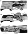
File:AnsonKarabinMartin1939 fig13-15.jpg ==Figs. 13 to 15. Reconstruction of the stapedial region in a fetus at term==(1,280 × 1,519 (171 KB)) - 08:46, 22 October 2017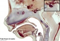
File:Fetal head section 02.jpg [[Category:Human Fetus]] [[Category:Week 12]] [[Category:Endocrine]] [[Category:Pituitary]](1,200 × 821 (171 KB)) - 12:43, 18 March 2012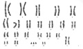
File:Trisomy21female.jpg ...ategory:Genetic Abnormalities]] [[Category:Human Embryo]] [[Category:Human Fetus]](480 × 284 (12 KB)) - 12:10, 20 July 2015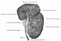
File:Bailey320.jpg --[[User:S8600021|Mark Hill]] 14:49, 27 May 2011 (EST) Fetus CRL 10cm should be about fertilization age 13 weeks or a gestational age (L(869 × 582 (75 KB)) - 18:08, 19 September 2011
File:Fetal head section 03.jpg [[Category:Human Fetus]] [[Category:Week 12]] [[Category:Endocrine]] [[Category:Pituitary]](1,200 × 821 (174 KB)) - 08:59, 28 March 2014
File:Jimenez-Castellanos1949 fig04-6.jpg ...menez-Castellanos1949 fig06.jpg|Fig. 6]]. Photomicrograph through a human fetus of 40 mm. at the middle level indicated in fig. 3. ac 15. (1) Suprarenal gl(735 × 1,695 (263 KB)) - 04:08, 17 August 2017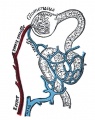
File:Gray1129.jpg ...ng to the state of secretion and the amount of fluid present in it. In the fetus and young subject the lining epithelial cells are polyhedral or even column(396 × 500 (55 KB)) - 11:39, 2 August 2019
File:Streeter028-30.jpg Lateral view of a model reconstructed from a human fetus 50 mm. crown-rump length (Carnegie Collection, No. 84). The cistern and the ...uction of the left membranous labyrinth and the periotic spaces in a human fetus 85 mm. crown-rump length (Carnegie Collection, No. 1400-30), enlarged 11.4(748 × 1,000 (134 KB)) - 17:30, 18 May 2015
File:Trisomy21male.jpg ...ategory:Genetic Abnormalities]] [[Category:Human Embryo]] [[Category:Human Fetus]](480 × 284 (10 KB)) - 12:28, 3 June 2018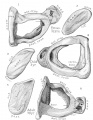
File:AnsonKarabinMartin1939 fig01-06.jpg 1, 2 - Fetus at term(1,280 × 1,650 (324 KB)) - 08:34, 22 October 2017
File:Fetal head section.jpg [[Category:Human Fetus]] [[Category:Week 12]] [[Category:Musculoskeletal]] [[Category:Tongue]] [[C(1,200 × 821 (167 KB)) - 08:09, 13 November 2018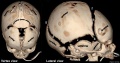
File:Skull CT normal sutures 01.jpg ..._human_fetus_of_43_millimeters_greatest_length|Historic - skull of a human fetus of 43 millimeters greatest length]] | [[Computed Tomography]](1,000 × 526 (89 KB)) - 23:13, 21 March 2018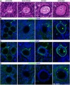
File:Human ovary follicle basement membrane 01.jpg [[Category:Human]][[Category:Human Fetus]][[Category:Fluorescence]][[Category:Ovary]][[Category:Oocyte]](660 × 800 (184 KB)) - 09:23, 1 May 2015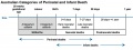
File:Australian categories perinatal and infant death graph.jpg ...hweight. The death is indicated by the fact that after such separation the fetus does not breathe or show any other evidence of life, such as beating of the(800 × 298 (43 KB)) - 18:19, 27 May 2018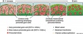
File:Model of human fetal ovarian cord development 01.jpg [[Category:Human]][[Category:Human Fetus]][[Category:Cartoon]][[Category:Ovary]][[Category:Oocyte]](800 × 330 (104 KB)) - 08:38, 1 May 2015
File:1 Min Embryo - Human timeline.mp4 ...y-human-timeline UNSW theBox] | [[BGDA Lecture - Development of the Embryo/Fetus 1|BGDA lecture]] | [[BGDA Practical - Fertilization to Implantation|BGDA Pr(5.28 MB) - 10:40, 28 April 2016


.jpg)


