Search results
From Embryology
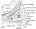
File:Keith1902 fig039.jpg ...atinous tissue is absorbed, so that the malleus and incus and developing {{stapes}}, with the chorda tympani, become surrounded by the hypoblasts lining of t(751 × 600 (114 KB)) - 19:35, 17 April 2018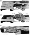
File:AnsonKarabinMartin1939 fig13-15.jpg In these and in the succeeding figures ant. crus. indicates anterior crus of stapes; cart. or cartil., cartilage; far. c., facial canal ; fen. cart, fenestral(1,280 × 1,519 (171 KB)) - 08:46, 22 October 2017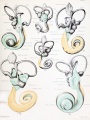
File:Streeter028-30.jpg * Impressio staped. - area in contact with base of stapes ...shown in green. Although the greater part of the cistern abuts against the stapes, it will be noted that it is also begiiming to spread over the liorsal surf(748 × 1,000 (134 KB)) - 17:30, 18 May 2015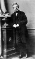
File:Karl Bogislaus Reichert.jpg ...#cartilage|cartilage]] band. The dorsal ends form the middle ear ossicle (stapes) and temporal bone styloid process, the ventral part ossifies to form hyoid(357 × 600 (40 KB)) - 17:31, 13 September 2016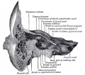
File:Gray0913.jpg ...ex border is upward. In the recent state it is occupied by the base of the stapes, the circumference of which is fixed by the annular ligament to the margin(671 × 600 (98 KB)) - 07:02, 19 August 2012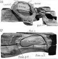
File:AnsonKarabinMartin1939 fig16-17.jpg In these and in the succeeding figures ant. crus. indicates anterior crus of stapes; cart. or cartil., cartilage; far. c., facial canal ; fen. cart, fenestral(1,280 × 1,301 (163 KB)) - 08:54, 22 October 2017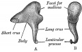
File:Gray0917.jpg ...lar process is tipped with cartilage, and articulates with the head of the stapes(600 × 379 (44 KB)) - 17:24, 18 May 2015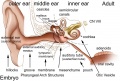
File:Adult hearing embryonic origins.jpg ** Stapes - Pharyngeal Arch 2 cartilage Neural crest (ectoderm)(1,000 × 675 (80 KB)) - 14:26, 8 May 2018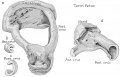
File:Anson1948 fig16.jpg ==Fig. 16. Drawings of a reconstruction of the stapes in a fetus at term==(1,280 × 817 (133 KB)) - 11:07, 14 October 2017
File:HansonAnsonBast1959 fig02.jpg ...be marle: the medial (mel larger Pllrt the lobe lI'I'lI gil'e rise 10 the stapes; the pm'llale"al to Ihe nerve wiLL become the latero- h!Jllle; Ihe iniercol(1,280 × 824 (130 KB)) - 14:14, 1 January 2018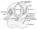
File:Keith1902 fig035.jpg ...lato-quadrate bar (cartilaginous skeleton of maxillary process), while the stapes is an independent formation developed round the stapedial artery. It may be(800 × 633 (125 KB)) - 12:46, 28 June 2016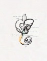
File:Streeter027.jpg * Impressio staped., area in contact with base of stapes(774 × 1,000 (51 KB)) - 17:31, 18 May 2015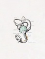
File:Streeter026.jpg * Impressio staped., area in contact with base of stapes(774 × 1,000 (45 KB)) - 17:31, 18 May 2015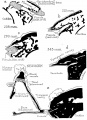
File:Anson1948 fig08.jpg ...the newborn (c), the capsule has acquired an adult appearance, as has the stapes.(1,280 × 1,764 (207 KB)) - 20:25, 16 October 2017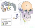
File:Cranial neural crest skeletal fate 01.jpg * '''ST''' - stapes(800 × 633 (59 KB)) - 22:36, 11 May 2019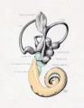
File:Streeter029.jpg * Impressio staped., area in contact with base of stapes(774 × 1,000 (74 KB)) - 17:32, 18 May 2015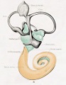
File:Streeter031.jpg * Impressio staped., area in contact with base of stapes(774 × 1,000 (79 KB)) - 17:34, 18 May 2015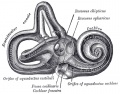
File:Gray0921.jpg ...is the fenestra vestibuli, closed, in the fresh state, by the base of the stapes and annular ligament. On its medial wall, at the forepart, is a small circu(640 × 500 (95 KB)) - 07:30, 19 August 2012
File:Gray0920.jpg ...is the fenestra vestibuli, closed, in the fresh state, by the base of the stapes and annular ligament. On its medial wall, at the forepart, is a small circu(600 × 438 (69 KB)) - 07:28, 19 August 2012
File:Gray0914.jpg ...ex border is upward. In the recent state it is occupied by the base of the stapes, the circumference of which is fixed by the annular ligament to the margin(708 × 500 (102 KB)) - 07:13, 19 August 2012


