Search results
From Embryology
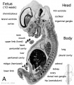
File:Human- fetal week 10 sagittal plane A.jpg ...he embryonic period (up to week 8) but still only 2 weeks into early fetal development.(500 × 573 (96 KB)) - 16:19, 27 April 2010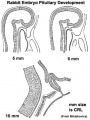
File:Pituitary rabbit development.jpg ==Rabbit Pituitary Development== Cartoon showing the changes in the embryonic rabbit pituitary.(374 × 500 (33 KB)) - 14:52, 27 May 2014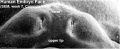
File:Stage19 em11.jpg ...EM images focussing on this developmentally important region and time for embryonic cleft lip and palate.(800 × 329 (46 KB)) - 10:11, 23 February 2014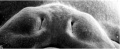
File:Stage19 em01.jpg ...EM images focussing on this developmentally important region and time for embryonic cleft lip and palate.(800 × 329 (37 KB)) - 08:17, 23 February 2014
File:Grasshopper lifecycle.jpg # As soon as the eggs are laid, they begin embryonic development and reach an advanced stage in which they enter diapause and pass the winte # In spring the eggs complete embryonic development and hatch.(1,072 × 814 (122 KB)) - 13:40, 16 February 2016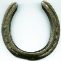
File:Horseshoe.jpg ...]] within the lateral plate [[mesoderm]] that forms during week 3 of human development. ...:''' [[Renal_System_-_Abnormalities|Renal Abnormalities]] | [[Renal System Development]](400 × 400 (32 KB)) - 09:09, 13 October 2016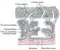
File:Gray0037.jpg ...evelopment contain core of mesoderm. Tertiary villi then have blood vessel development within this core. Extra-embryonic mesoderm grows into villi, covers the entire surface of chorionic sac.(500 × 412 (74 KB)) - 11:21, 9 June 2014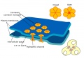
File:Gap junction 01.jpg * Also in embryonic development (see [[Blastocyst Development]])(800 × 562 (69 KB)) - 12:35, 25 March 2015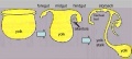
File:Endoderm cartoon.jpg ==Cartoon of endoderm development== ...3 images is from the animation [[Development_Animation_-_Endoderm|Endoderm Development]](587 × 262 (31 KB)) - 19:42, 11 June 2013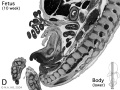
File:Human- fetal week 10 lower body D.jpg ...he embryonic period (up to week 8) but still only 2 weeks into early fetal development.(600 × 450 (91 KB)) - 15:36, 27 April 2010
File:Stages 1-5 mouse.pdf Table 1: Mouse embryonic stages of development from fertilization to zona free blastocyst (stages 1 to 5).(29 KB) - 13:42, 31 March 2012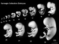
File:Human Carnegie stage 10-23.jpg ==Human Embryonic Development== ...tion, embryos compiled and scaled from original images. Note that weeks of development are form fertilisation (fertilization age), not gestational age {{GA}} that(1,024 × 768 (95 KB)) - 11:38, 16 February 2018
File:Elephant and calf.jpg * 50 days - embryonic vesicle earliest observation * 71 days - embryonic heartbeat and allantois visible as a single sacculation(538 × 404 (52 KB)) - 20:24, 1 November 2013
File:Morphological differences in early mouse embryonic development.png ...hological and lineage specification steps during the early mouse embryonic development== ...is formed as a layer separating epiblast from blastocoel (E4.5). After 4.5 embryonic days, the preimplantation embryo contains more than 100 cells.(2,061 × 886 (455 KB)) - 12:47, 18 August 2016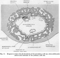
File:Hamilton1949 fig12.jpg ==Fig.12. Diagram to show the development of the secondary yolk sac, extra-embryonic coelom and remnant of the primary yolk sac==(1,312 × 1,265 (267 KB)) - 15:30, 15 May 2018
File:Hertig1956 fig82.jpg ...ssible to say what effect, if any, such an abnormality would have upon the development of the body stalk and the embryo. [[:Category:Carnegie Embryo 8299|Carnegie(1,000 × 796 (181 KB)) - 14:37, 25 February 2017
File:Human- fetal week 10 sagittal plane B.jpg ...he embryonic period (up to week 8) but still only 2 weeks into early fetal development.(500 × 573 (99 KB)) - 15:49, 27 April 2010
File:Bat - neural development 01.jpg ==Bat neural Development (stage 14)== :'''Links:''' [[Bat Development]] | [[Neural System Development]] | [[Carnegie stage 14]](733 × 498 (33 KB)) - 13:17, 6 July 2012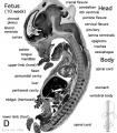
File:Human- fetal week 10 sagittal plane D.jpg ...he embryonic period (up to week 8) but still only 2 weeks into early fetal development.(500 × 573 (105 KB)) - 07:41, 1 May 2011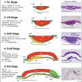
File:Cattle embryo staging 01.jpg ...al sections (H&E or CER1 stained) of, cattle embryos at the five stages of development between hatching and the start of {{gastrulation}}, based on data from sect ...ural) hypoblast; PS, primitive streak; RL, Rauber’s Layer (polar TB); VH, (embryonic) visceral hypoblast. All bars are 100 μm.(1,961 × 1,962 (708 KB)) - 13:45, 22 November 2018


