Search results
From Embryology
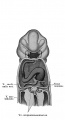
File:Keibel Mall 2 425.jpg After W. His, Anat. mensch. Embry., iii, Leipzig, 1885, p. 1S6, Fig. 119, and Atlas(745 × 1,376 (97 KB)) - 19:25, 14 January 2017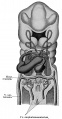
File:Keibel Mall 2 426.jpg (After W. His, Anat. mensch. Embry., iii, Leipzig, 1885, p. 1S6, Fig. 119, and Atlas(715 × 1,376 (150 KB)) - 19:25, 14 January 2017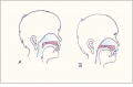
File:Normal and Cleft Palate.JPG ...d on Gray's Anatomy for Students <ref> Drake, R., Vogl, A. W., Mitchel, A. W. M. (2010). Gray's Anatomy for Students (Ed. 2). Elsevier, Churchil Livings(781 × 512 (29 KB)) - 19:52, 5 October 2011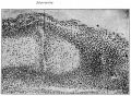
File:Bailey153.jpg Photograph from preparation by Dr. W. C. Clarke.(799 × 585 (175 KB)) - 12:01, 18 January 2011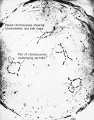
File:Rugh 042.jpg Courtesy, W. R. Duryee, Laboratory of Terrestrial Magnetism, Washington, D. C.(627 × 800 (122 KB)) - 09:03, 12 April 2013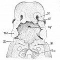
File:Minot1897 fig082.jpg .... Lit, Lung. Md, Mandible. II, Second branchial arch. X I0 diams. — (After W. His.)(697 × 692 (124 KB)) - 15:32, 7 April 2014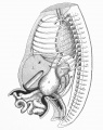
File:Mall1908a fig6a.jpg X 8 times. 1-13, dorsal ganglia; SB, suprarenal body: S, stomach; C, caecum; W, Wolffian body; K, kidney; L, liver.(1,280 × 1,623 (388 KB)) - 11:34, 29 July 2018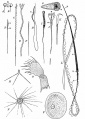
File:Bailey005.jpg k, End knob; w, middle piece; u, undulatory membrane.(747 × 1,050 (138 KB)) - 11:27, 21 May 2020
File:Gray1110.jpg ...ary or testis is formed. ug. Sinus urogenitalis. W. Left Wolffian body. w, w. Right and left Wolffian ducts. ...rus. The uterine tube of the right side is marked m. v. Vulva. va. Vagina. W. Scattered remains of Wolffian tubes near it (paroöphoron of Waldeyer).(414 × 1,200 (86 KB)) - 10:48, 5 June 2013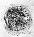
File:Hamilton1943 fig04.jpg ...pper right-hand corner of the photograph globular detritus is seen. x 480. w..H.(1,022 × 1,101 (108 KB)) - 21:24, 30 October 2017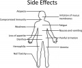
File:Chemotherapy Side Effects.jpg Licensed content author - Joseph W. Nichols,You Han Bae Nichols, J. W., & Bae, Y. H. (2012). Odyssey of a cancer nanoparticle: from injection sit(1,050 × 866 (127 KB)) - 12:25, 23 October 2015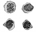
File:Keibel Mall 2 362.jpg ...multiply by mitotic division of the lymph-glands was first demonstrated by W. Flemming.(674 × 600 (52 KB)) - 11:02, 29 March 2014
File:Gray1110b.jpg ...rus. The uterine tube of the right side is marked m. v. Vulva. va. Vagina. W. Scattered remains of Wolffian tubes near it (paroöphoron of Waldeyer).(578 × 600 (44 KB)) - 10:35, 5 June 2013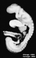
File:Wen1928-Fig01c.jpg ...X 16.6. Embryo H984 (fig. 1, C) was donated to the collection by Drs. E. W. Rawson and B. H. Foreman of Tacoma, Washington.(398 × 641 (28 KB)) - 21:30, 21 April 2016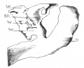
File:Odgers1930 fig03.jpg ...in an 11.4 mm. embryo. D. duodenum; B.D. bile-duct; V.P. ventral pancreas; W and W1 mark the two ventral pancreatic ducts.(926 × 755 (122 KB)) - 12:03, 30 June 2015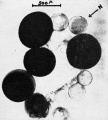
File:Rugh 038.jpg Courtesy, W. R. Duryee, 1950, Ann. N. Y. Acad. ScL, 50, Art. 8.(721 × 800 (107 KB)) - 08:40, 12 April 2013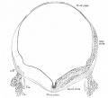
File:Bailey401.jpg a. W., Spinal V; V. s., Gasserian ganglion; V. m., part of efferent root of V ne(788 × 718 (61 KB)) - 00:54, 14 February 2011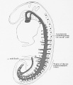
File:Keibel Mall 2 414.jpg (NT. 7). After W. Felix, 1910.)(1,000 × 1,162 (132 KB)) - 11:34, 15 January 2017
File:ZUltrasound System.jpg Image Author: Daniel W. Rickey(262 × 400 (49 KB)) - 14:24, 19 August 2014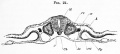
File:Foster024.jpg M. medullary canal ; Pv. mesoblastic somite ; w. rudiment of Wolffian duct; A. epiblast; C. hypoblast ; Ch. notochord ; Ao.(839 × 380 (54 KB)) - 07:27, 9 January 2011


