Search results
From Embryology
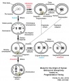
File:Aneuploidy model based on fragmentation.jpg ==Model for the Origin of Human Embryonic Aneuploidy Based on Fragmentation Timing== ...omal abnormalities and undergoes fragmentation as a survival mechanism. As development proceeds, these fragments either remain or are reabsorbed by the blastomere(668 × 790 (105 KB)) - 13:44, 1 April 2019
File:Bat embryo stage 10-13.jpg ==Bat Embryonic Development (stage 10-13)== :'''Links:''' [[Bat Development]] | [[:File:Bat embryo stage 10-13.jpg|stage 10 to 13]] | [[:File:Bat embry(547 × 767 (23 KB)) - 01:34, 29 April 2011
File:ThyroidDevelopment.png This image summarises the endodermal and mesodermal contribution to the development of the thyroid gland. The progenitor cells are from anterior endoderm and r ...ed to understand what has gone before, you have not included much in fetal development. There is no explanation of the molecular factors shown in the figure.(1,996 × 1,212 (49 KB)) - 11:46, 9 November 2014
File:Human blastocyst day 3-6.mov ...asive imaging of human embryos before embryonic genome activation predicts development to the blastocyst stage. ...asive imaging of human embryos before embryonic genome activation predicts development to the blastocyst stage(4.26 MB) - 16:08, 15 October 2010
File:Embryonic upper limb - brachial and superficial brachial artery.jpg ==Embryonic upper limb - brachial and superficial brachial artery == ...development of the brachial artery and the superficial brachial artery in embryonic upper limb.(1,280 × 314 (122 KB)) - 14:25, 2 February 2020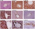
File:Histology of human embryonic liver at 11 weeks.png ==Histology of human embryonic liver at 11 weeks== Paraffin-embedded sections of human embryonic liver at 11 weeks (9 weeks of gestation) stained for Hematoxylin and Eosin(1,988 × 1,642 (4.17 MB)) - 22:02, 8 November 2014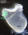
File:Stage9 bf2-primordial germ cell region.jpg # the relative size of the embryo and the associated extra-embryonic coeloms. # the shape of the early folded embryonic disc and rostro-caudal bendings.(814 × 1,000 (72 KB)) - 21:20, 17 May 2015
File:Bailey061.jpg ...ys and 22 hours after insemination (younger than B but further advanced in development), showing beginning of proamniotic cavity. ...astocyst 8 days after insemination (younger than B but further advanced in development), showing more advanced proamniotic cavity.(878 × 1,086 (184 KB)) - 15:02, 26 January 2011
File:1 Min Embryo - Human timeline.mp4 ...mplantation to 8 Weeks|BGDA Prac 6]] | [[Embryonic Development]] | [[Fetal Development]] | [[One Minute Embryology]] Lets look at an overview of human development(5.28 MB) - 10:40, 28 April 2016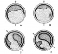
File:Rugh 077.jpg ...r V. Hamburger and B. Mayer, unpublished. Redrawn from Spemann: "Embryonic Development and Induction," New Haven, Yale University Press.(861 × 800 (171 KB)) - 13:43, 12 April 2013
File:Human- fetal week 10 sagittal plane A.jpg ...he embryonic period (up to week 8) but still only 2 weeks into early fetal development.(500 × 573 (96 KB)) - 16:19, 27 April 2010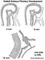
File:Pituitary rabbit development.jpg ==Rabbit Pituitary Development== Cartoon showing the changes in the embryonic rabbit pituitary.(374 × 500 (33 KB)) - 14:52, 27 May 2014
File:Stage19 em11.jpg ...EM images focussing on this developmentally important region and time for embryonic cleft lip and palate.(800 × 329 (46 KB)) - 10:11, 23 February 2014
File:Stage19 em01.jpg ...EM images focussing on this developmentally important region and time for embryonic cleft lip and palate.(800 × 329 (37 KB)) - 08:17, 23 February 2014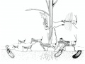
File:Grasshopper lifecycle.jpg # As soon as the eggs are laid, they begin embryonic development and reach an advanced stage in which they enter diapause and pass the winte # In spring the eggs complete embryonic development and hatch.(1,072 × 814 (122 KB)) - 13:40, 16 February 2016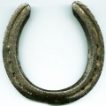
File:Horseshoe.jpg ...]] within the lateral plate [[mesoderm]] that forms during week 3 of human development. ...:''' [[Renal_System_-_Abnormalities|Renal Abnormalities]] | [[Renal System Development]](400 × 400 (32 KB)) - 09:09, 13 October 2016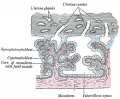
File:Gray0037.jpg ...evelopment contain core of mesoderm. Tertiary villi then have blood vessel development within this core. Extra-embryonic mesoderm grows into villi, covers the entire surface of chorionic sac.(500 × 412 (74 KB)) - 11:21, 9 June 2014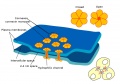
File:Gap junction 01.jpg * Also in embryonic development (see [[Blastocyst Development]])(800 × 562 (69 KB)) - 12:35, 25 March 2015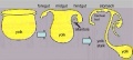
File:Endoderm cartoon.jpg ==Cartoon of endoderm development== ...3 images is from the animation [[Development_Animation_-_Endoderm|Endoderm Development]](587 × 262 (31 KB)) - 19:42, 11 June 2013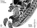
File:Human- fetal week 10 lower body D.jpg ...he embryonic period (up to week 8) but still only 2 weeks into early fetal development.(600 × 450 (91 KB)) - 15:36, 27 April 2010


