Search results
From Embryology
File:Bailey402.jpg [[Category:Cranial Nerve]](640 × 483 (116 KB)) - 08:42, 23 June 2015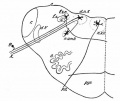
File:Bailey369.jpg ...ection through the lower human medulla showing the origin of the X and XII cranial nerves== [[Category:Neural]][[Category:Cranial Nerve]](529 × 446 (33 KB)) - 08:15, 16 February 2016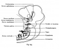
File:Kollmann637.jpg ==Fig. 637. The Urspnias folseadea the cranial nerves is shown== [[Category:Cranial Nerve]](679 × 561 (51 KB)) - 23:06, 4 September 2014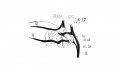
File:Lisser1911 fig17.jpg ==Fig. 17.Graphic reconstruction of 9th, 10th, and 12th cranial nerves in larynx region of human Embryo no. 43== ...l and inferior laryngeal nerves; m br., motor branch of superior laryngeal nerve.(1,581 × 916 (80 KB)) - 18:15, 15 June 2016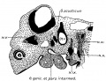
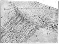
File:Streeter1906 fig06.jpg In Fig. 6 the ventral or motor division of the facial nerve can be seen cut obliquely at the ventral edge of the geniculate ganglion. I [[Category:Neural]][[Category:Cranial Nerve]](1,089 × 833 (240 KB)) - 13:31, 7 December 2016
File:Frazer1928 plate01.jpg Top, cranial slope of hind-brain and sagittal section of hind- and mid-brain in 12 mm. e N. decussation of fourth nerve; a.l. alar lamina; b.l. basal lamina; f.l. floor lamina; t. tectum of mid-(1,442 × 1,827 (428 KB)) - 12:58, 9 January 2017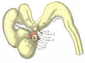
File:Gilbert1957 fig14.jpg ...y the same age and size. The relation of the principal branches of cranial nerve V to the four peripheral condensations into which the four rectus muscles g ...c vesicle, into which the primordia of the four rectus muscles have grown. Cranial nerves III, IV, and VI have reached their respective eye-muscle primordia:(1,280 × 947 (218 KB)) - 15:52, 24 May 2017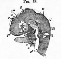
File:Foster038.jpg ...diments of the fifth cranial nerve, VII. of the seventh. Below the seventh nerve is seen the auditory vesicle b. The head having been subjected to pressure,(432 × 416 (46 KB)) - 13:14, 29 January 2016
File:Wheeler024.jpg Those cranial nerves which were identified are shown by lines. Only the first spinal nerve is shown.(592 × 700 (49 KB)) - 09:11, 16 February 2011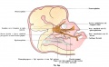
File:Kollmann639.jpg from the main stems of the nerve ganglion hervorgelienden First the ophthalmic nerve, ciliary ganglion to the top of the bulb.(1,068 × 655 (99 KB)) - 23:07, 4 September 2014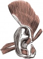
File:Gray0906.jpg ...erge to be inserted by a thin, flattened tendon into the upper part of the cranial surface of the auricula. ...by short aponeurotic fibers. They are inserted into the lower part of the cranial surface of the concha.(438 × 600 (81 KB)) - 06:28, 19 August 2012
File:Gilbert1957 plate04.jpg ...y the same age and size. The relation of the principal branches of cranial nerve V to the four peripheral condensations into which the four rectus muscles g ...c vesicle, into which the primordia of the four rectus muscles have grown. Cranial nerves III, IV, and VI have reached their respective eye-muscle primordia:(2,165 × 2,232 (694 KB)) - 09:10, 2 January 2018
File:Stage 22 image 219.jpg * '''Neural''' - cranial nerve 8 (CNVIII), brain stem region, ventricular space and choroid plexuses.(1,250 × 892 (295 KB)) - 11:42, 14 June 2016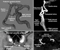
File:Trigeminal artery 01.jpg [[Category:Cranial Nerve]][[Category:Artery]](947 × 800 (102 KB)) - 23:48, 8 September 2019
File:Macklin1914 plate06.jpg ...hatica and the fenestrae for the vestibular division of the eighth cranial nerve. It contains the first, or unwound, portion of the ductus cochlearis. I sha The medial surface (fig. 7) is more extensive than the lateral. Its cranial and dorsal borders are the same as those of the lateral surface; its ventra(1,928 × 2,837 (567 KB)) - 22:59, 20 June 2016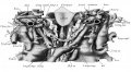
File:Thyng1914 plate6.jpg * Plate 6 shows some of the cranial nerves on both sides. * The superior laryngeal nerve (N.Ls., pi. 3; N.laryng.s., pi. 6) arises at the ganglion nodosum, slightly(2,000 × 1,098 (422 KB)) - 14:19, 18 May 2014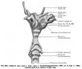
File:Keibel Mall 308.jpg ...of the nasal septum and gives rise to parts of the membranous floor of the cranial cavity and the roof of the mouth ([[:File:Keibel_Mall_310.jpg|Fig. 310]]).(677 × 578 (43 KB)) - 13:18, 27 January 2014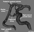
File:Trigeminal artery 02.jpg [[Category:Blood Vessel]][[Category:Neural]][[Category:Cranial Nerve]](520 × 490 (35 KB)) - 23:49, 8 September 2019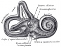
File:Gray0921.jpg ...which ends in a cul-de-sac between the layers of the dura mater within the cranial cavity. On the upper wall or roof is a transversely oval depression, the re(640 × 500 (95 KB)) - 07:30, 19 August 2012
File:Gray0920.jpg ...which ends in a cul-de-sac between the layers of the dura mater within the cranial cavity. On the upper wall or roof is a transversely oval depression, the re(600 × 438 (69 KB)) - 07:28, 19 August 2012


