Search results
From Embryology
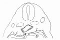
File:Flint1906 textfig01.jpg C = Coelom. SV = Sinus venosus. VM = Mesocardium posterior.(700 × 473 (33 KB)) - 14:21, 8 April 2020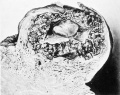
File:Mall1916 fig03.jpg The embryo lies within the coelom, and bands of -magma fibrils radiate from the amnion to the chorionic wall.(995 × 787 (145 KB)) - 10:51, 22 April 2014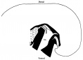
File:Wyburn1939-text-fig05.jpg ...blood vessels. Black = mesoderm. Small dots = gut. U .C. = umbilical cord coelom. Black line indicates the plane of the umbilical veins.(1,300 × 935 (66 KB)) - 13:19, 15 September 2015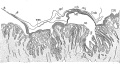
File:Minot1889 fig01.jpg ...doderm only; vt, vena terminals; mrs, mesoderm; sol, splanchnopleure; coe, coelom; nch, notochord; Md, medullary groove; my, myotome; Endo, endoderm; Ecto, e(1,200 × 644 (307 KB)) - 12:17, 4 April 2014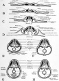
File:Patten054.jpg ...grams of cross sections at various stages to show the establishment of the coelom and mesenteries== ...all in the yolk-stalk region, results in the embryonic and extra-embryonic coelom retaining their open communication at this point for a long time after they(764 × 1,060 (179 KB)) - 09:11, 29 July 2011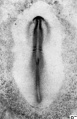
File:HHstage8-.jpg | 1-3 somites; coelom(727 × 1,125 (90 KB)) - 16:22, 2 June 2017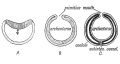
File:Keith1921 fig038.jpg '''C.''' Origin of mesoderm (black) and coelom from margin of primitive mouth, with formation of a ventral mesentery round(1,200 × 636 (118 KB)) - 11:40, 23 December 2014
File:Hertig1946b fig09b.jpg ...he communication between the exocoelomic space around the yolk-sac and the coelom (body cavity) within the embryo. Carnegie {{CE6488}}, sequence 2, X15. 104(800 × 586 (26 KB)) - 17:35, 7 August 2017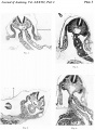
File:Wyburn1939-plate01.jpg A. = amnion. L.T. = lateral tissue plate. U. = umbilical vein. C. = coelom. Description in text.(1,600 × 2,180 (696 KB)) - 17:17, 14 September 2015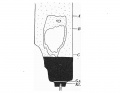
File:Wyburn1937 text-fig10.jpg ...cranial attachment of body stalk; between B and C is the extension of the coelom occupied by the midgut loop; D = the caudal attachment of the stalk; G.t. =(803 × 620 (40 KB)) - 17:53, 15 August 2015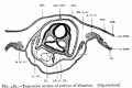
File:Bailey483.jpg ...nt at which extraembryonic body cavity passes over into intraembryonic (or coelom).(753 × 499 (64 KB)) - 08:47, 3 February 2011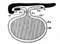
File:Bailey485.jpg ...f yolk sac; hs., parietal layer of yolk sac; hn., dermal umbilicus; lh l , coelom; lh 2 , exoccelom; m., mouth; st., yolk stalk.(583 × 425 (57 KB)) - 08:57, 3 February 2011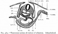
File:Bailey482.jpg ...t which extraembryonic body cavity passes over into the intraembryonic (or coelom proper).(709 × 419 (57 KB)) - 16:23, 9 August 2012
File:Horseshoe.jpg ...to describe the shape of the [[Coelomic Cavity Development|intra-embryonic coelom]] within the lateral plate [[mesoderm]] that forms during week 3 of human d(400 × 400 (32 KB)) - 09:09, 13 October 2016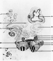
File:Shaw1932 fig03.jpg A, cerebral vesicle; B, heart; 0, coelom; D, intestine; E, primitive kidney; F, limb bud; G, dorsal aorta; H, neural(900 × 1,041 (81 KB)) - 17:09, 15 November 2017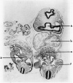
File:Shaw1932 fig04.jpg A, cerebral vesicle; B, heart; 0, coelom; D, intestine; E, primitive kidney; F, limb bud; G, dorsal aorta; H, neural(900 × 1,021 (100 KB)) - 17:09, 15 November 2017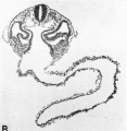
File:Hertig1946b fig10b.jpg ...of the embryo. The large spaces on either side of the gut are those of the coelom or body-cavity of the embryo. Carnegie {{CE6344}}, section 3-7-11, X75.(800 × 818 (85 KB)) - 08:37, 8 August 2017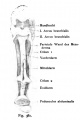
File:Kollmann381.jpg The intestinal system still consists of a straight tube of the Coelom surrounding the elongated cylindrical body, heart and runs Yolk sac are rem(463 × 695 (31 KB)) - 12:07, 20 October 2011
File:Flint1906 textfig02.jpg Through the pulmonary anlage. C = Coelom. PA = Pulmonary anlage.(671 × 520 (37 KB)) - 13:35, 8 April 2020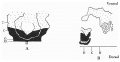
File:Wyburn1937 text-fig06.jpg ...ion 910; C = section 954; interval between B and 0 is the extension of the coelom into the stalk and occupied by the midgut loop. D = section 1002 at the lev(1,021 × 528 (55 KB)) - 17:44, 15 August 2015


