File list
From Embryology
This special page shows all uploaded files.
| Date | Name | Thumbnail | Size | User | Description | Versions |
|---|---|---|---|---|---|---|
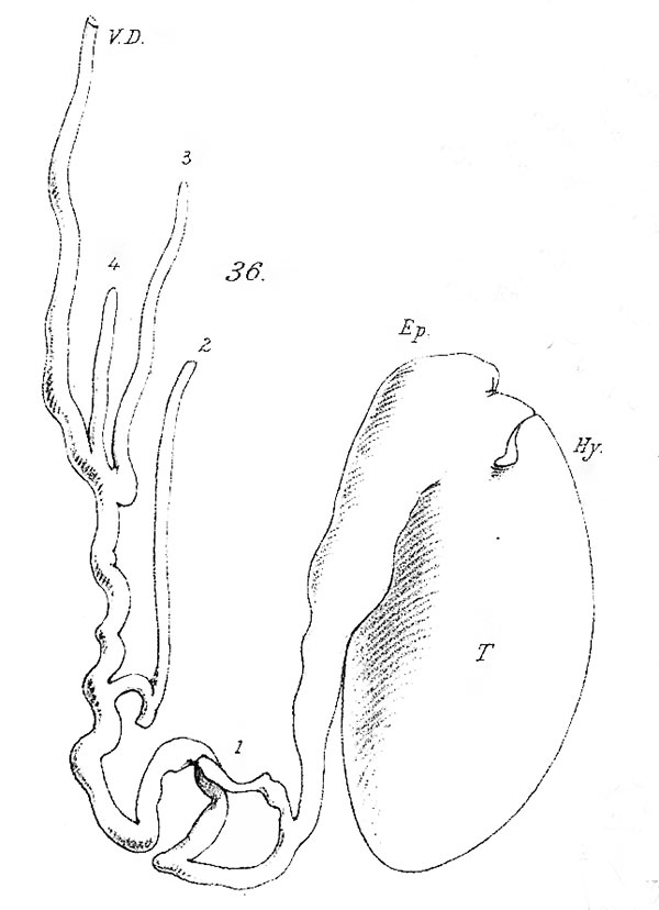
| 19:34, 14 April 2020 | Lockwood1887b fig36.jpg (file) |  |
53 KB | Z8600021 | 1 | |
| 18:19, 14 April 2020 | Lockwood1888b fig52.jpg (file) |  |
60 KB | Z8600021 | resize W 800px | 3 |
| 18:10, 14 April 2020 | Lockwood1888b fig49.jpg (file) |  |
69 KB | Z8600021 | adjust size W 800px | 3 |
| 18:04, 14 April 2020 | Lockwood1888b fig51.jpg (file) |  |
88 KB | Z8600021 | resize W 800px | 3 |
| 14:28, 14 April 2020 | Lockwood1888a fig47.jpg (file) |  |
279 KB | Z8600021 | resize | 3 |
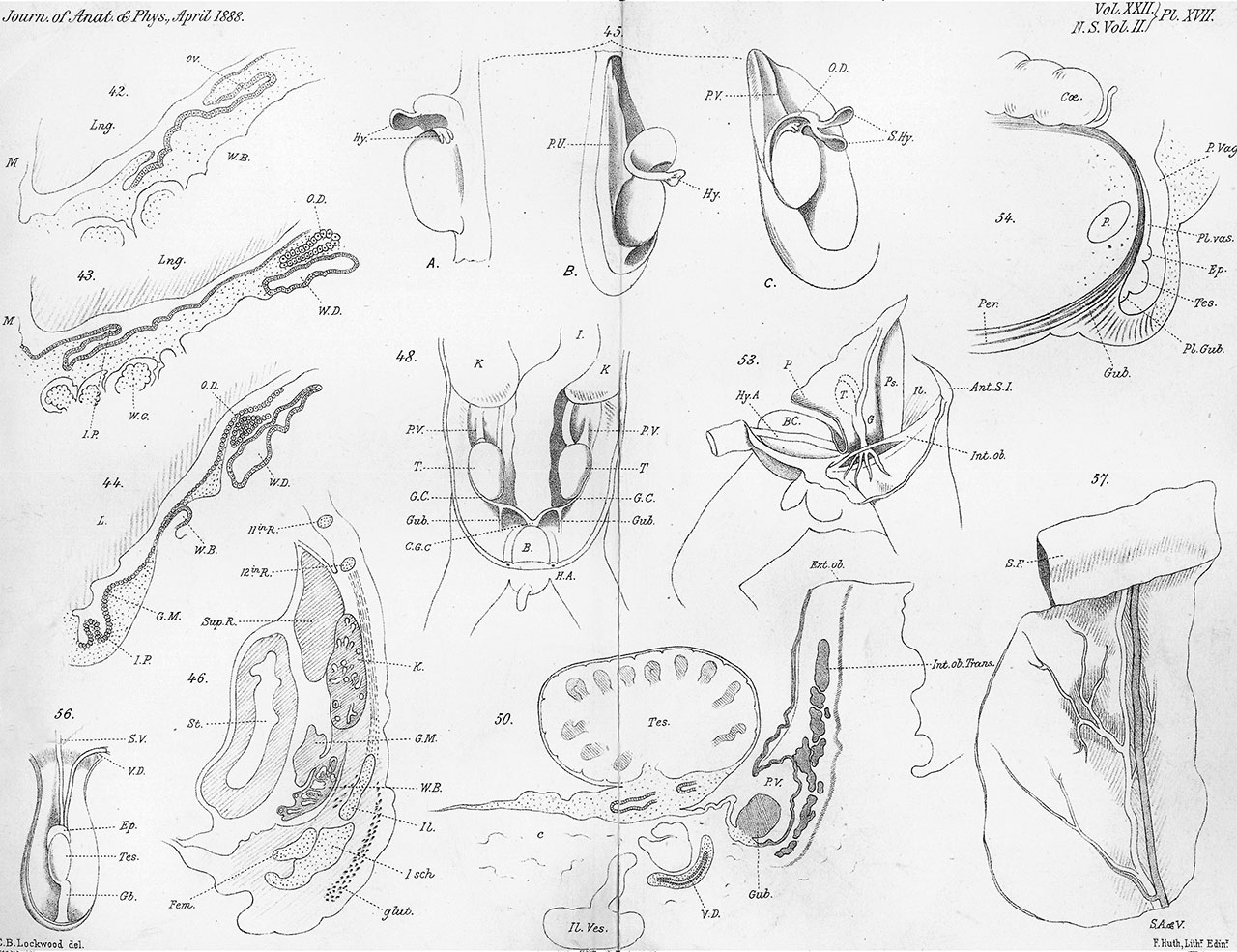
| 14:11, 14 April 2020 | Lockwood1888a plate07.jpg (file) |  |
285 KB | Z8600021 | scale W 1280px | 3 |
| 13:50, 14 April 2020 | Lockwood1887b fig41.jpg (file) |  |
129 KB | Z8600021 | contrast and size | 2 |
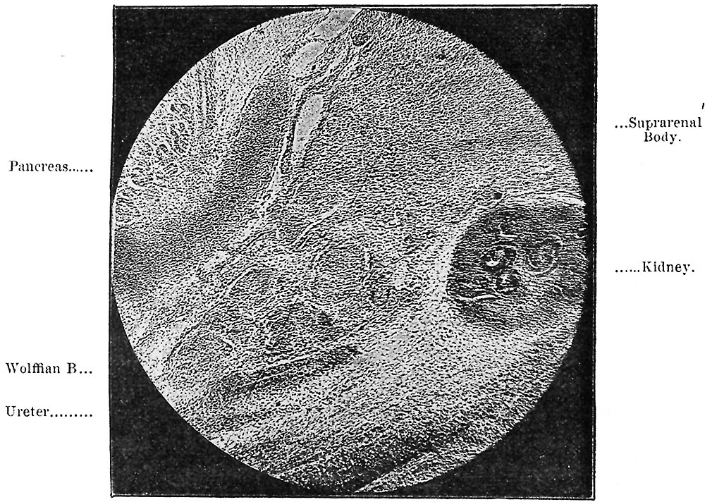
| 12:58, 14 April 2020 | Lockwood1887b fig38.jpg (file) |  |
65 KB | Z8600021 | adjust size contrast | 2 |
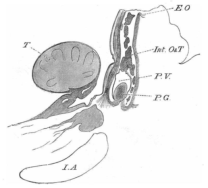
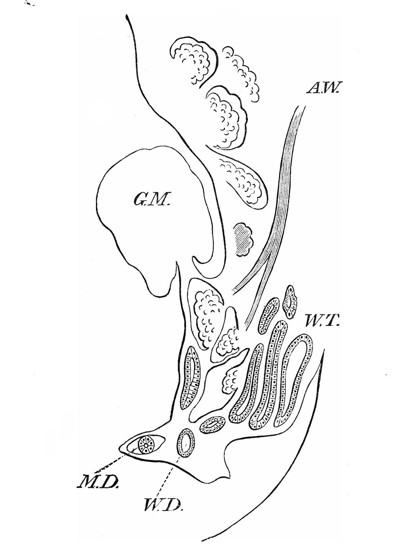
| 23:29, 13 April 2020 | Lockwood1887b fig39.jpg (file) |  |
93 KB | Z8600021 | 2 | |
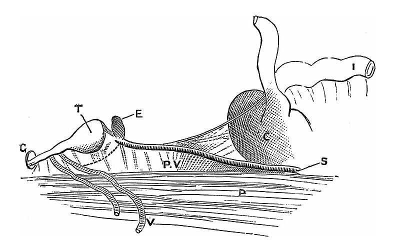
| 23:18, 13 April 2020 | Lockwood1887b fig40.jpg (file) |  |
92 KB | Z8600021 | 2 | |
| 23:02, 13 April 2020 | Lockwood1887b plate02.jpg (file) |  |
1.21 MB | Z8600021 | contrast | 2 |
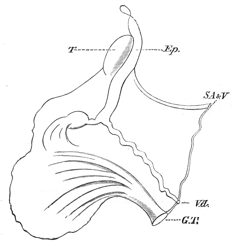
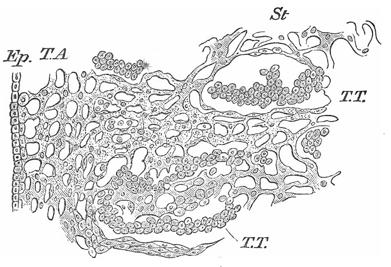
| 23:00, 13 April 2020 | Lockwood1887b fig37.jpg (file) |  |
102 KB | Z8600021 | adjust size contrast | 2 |
| 11:31, 9 April 2020 | 1906 A Text-book of Histology - Arranged Upon an Embryological Basis.pdf (file) |  |
16.15 MB | Z8600021 | Category:PDFCategory:HistologyCategory:Historic EmbryologyCategory:1900's | 1 |
| 11:31, 9 April 2020 | 1913 A Text-book of Histology - Arranged Upon an Embryological Basis.pdf (file) | |
19.56 MB | Z8600021 | Category:PDFCategory:HistologyCategory:Historic EmbryologyCategory:1910's | 1 |
| 15:17, 8 April 2020 | Flint1906 plate01.jpg (file) |  |
149 KB | Z8600021 | adjust size, sharpness | 3 |
| 14:35, 8 April 2020 | Flint1906 textfig02.jpg (file) |  |
37 KB | Z8600021 | ==Text Fig. 2. Section of embryo pig 3.5 mm long== Through the pulmonary anlage. C = Coelom. PA = Pulmonary anlage. {{Ref-Flint1906}} {{footer}} | 1 |
| 14:35, 8 April 2020 | Flint1906 textfig01.jpg (file) |  |
33 KB | Z8600021 | ==Text Fig. 1. Section of embryo pig 3.5 mm long== Showing head gut in the region of the upper part of the Mesocardium posterior. C = Coelom. SV = Sinus venosus. VM = Mesocardium posterior. {{Ref-Flint1906}} {{footer}} | 1 |
| 14:02, 8 April 2020 | Flint1906 fig01.jpg (file) |  |
33 KB | Z8600021 | 2 | |
| 14:02, 8 April 2020 | Flint1906 fig02.jpg (file) |  |
37 KB | Z8600021 | 2 | |
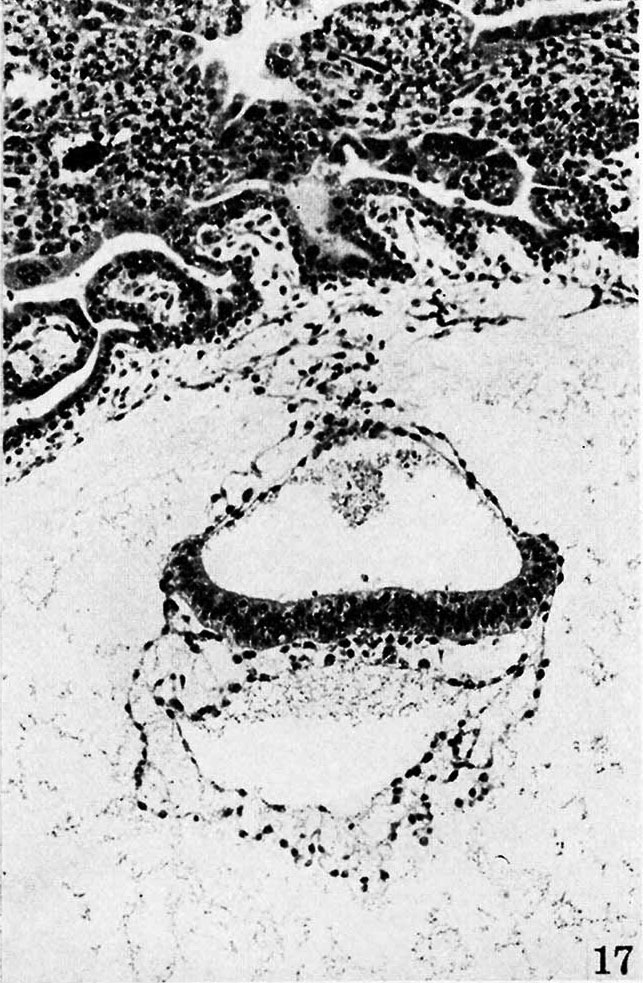
| 10:10, 8 April 2020 | Hertig1945d fig17.jpg (file) |  |
181 KB | Z8600021 | 1 | |
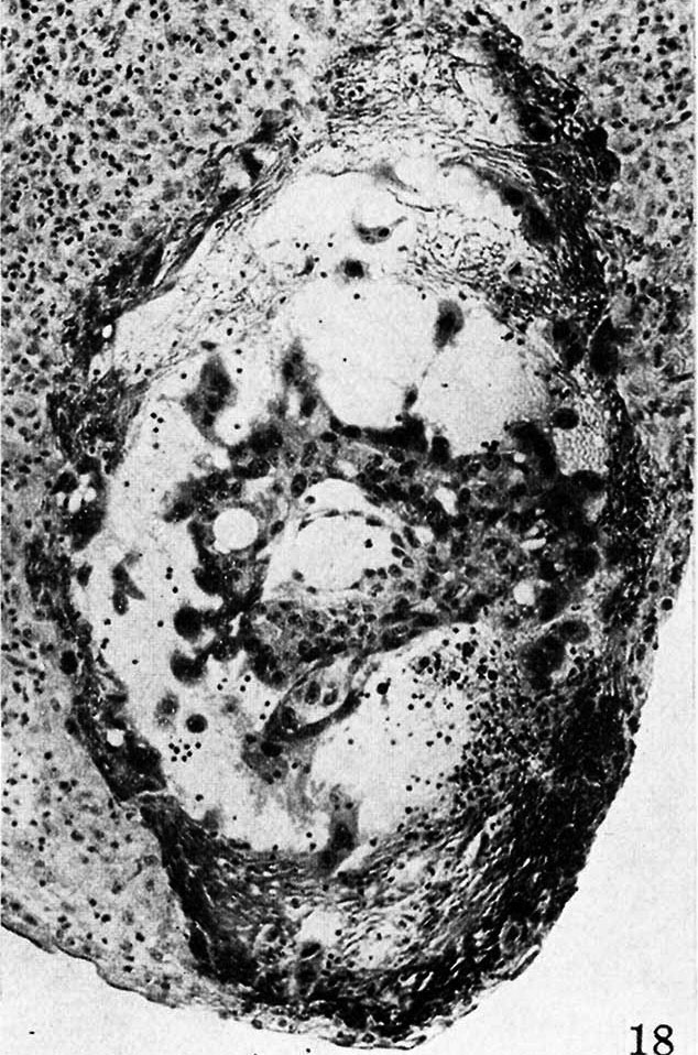
| 10:10, 8 April 2020 | Hertig1945d fig18.jpg (file) |  |
191 KB | Z8600021 | 1 | |
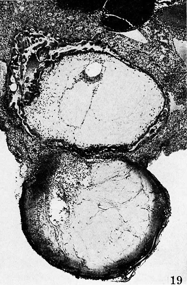
| 10:10, 8 April 2020 | Hertig1945d fig19.jpg (file) |  |
192 KB | Z8600021 | 1 | |
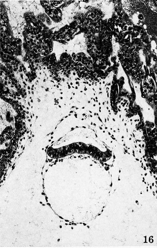
| 10:09, 8 April 2020 | Hertig1945d fig16.jpg (file) |  |
167 KB | Z8600021 | 1 | |
| 16:29, 5 April 2020 | Ferret.png (file) |  |
629 KB | Z8600021 | Category:Ferret | 1 |
| 11:24, 27 March 2020 | William James Hamilton.jpg (file) |  |
178 KB | Z8600021 | resize and adjust contrast | 2 |
| 09:56, 26 March 2020 | Theodor Heinrich Boveri.jpg (file) |  |
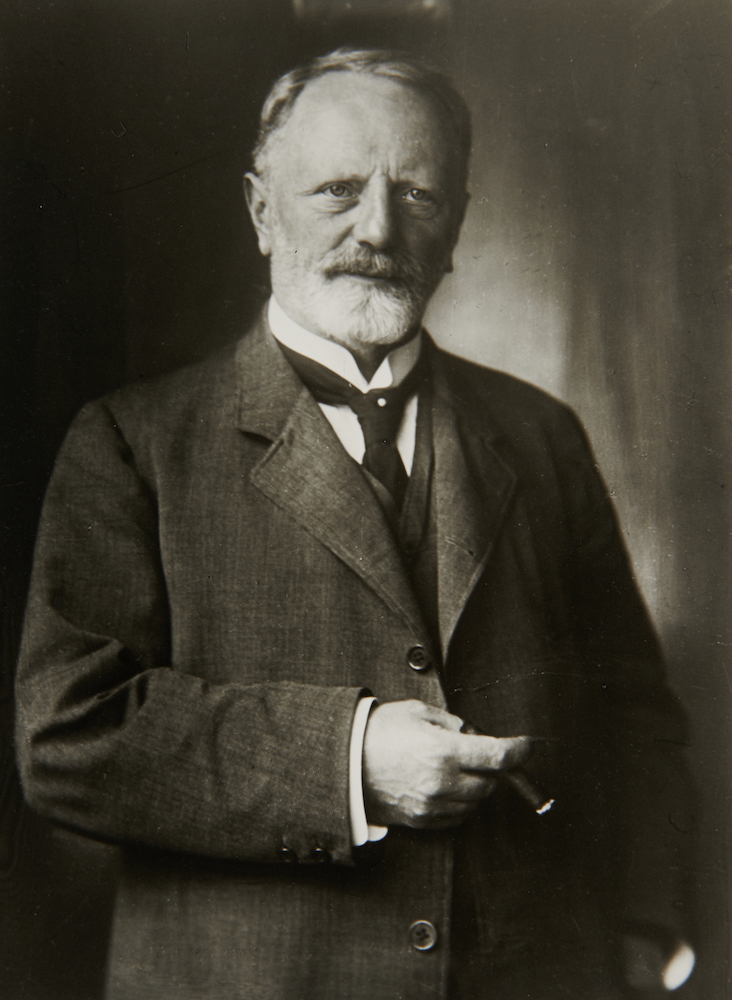
202 KB | Z8600021 | ==Theodor Boveri in 1908== ==Theodor Heinrich Boveri (1862 – 1915)== Theodor Heinrich Boveri (1862 – 1915) was a German biologist at the Institute of Zoology in Würzburg. In 1902 using the sea urchin model, identified the role of nuclear chromosomes in development. :Links: Embryologists ===Reference=== Unknown author - Theodor Boveri. In: Hugo Freund und Alexander Berg (Hrsg.): Geschichte der Mikroskopie. Leben und Werk großer Forscher. Bd. 1, Biologie, Umsc... | 1 |
| 11:56, 25 March 2020 | McMurrich1930 fig04.jpg (file) |  |
649 KB | Z8600021 | 2 | |
| 11:46, 25 March 2020 | McMurrich1930 fig03.jpg (file) |  |
582 KB | Z8600021 | 2 | |
| 11:42, 25 March 2020 | McMurrich1930 fig02.jpg (file) |  |
619 KB | Z8600021 | 2 | |
| 11:39, 25 March 2020 | McMurrich1930 fig01.jpg (file) |  |
720 KB | Z8600021 | 2 | |
| 11:15, 25 March 2020 | McMurrich1930 frontispiece.jpg (file) |  |
554 KB | Z8600021 | BW and scaled to 1280 pixels wide | 2 |
| 10:56, 25 March 2020 | McMurrich1930 fig87.jpg (file) |  |
267 KB | Z8600021 | BW and scaled to 1280 pixels wide | 2 |
| 10:53, 25 March 2020 | McMurrich1930 fig86.jpg (file) |  |
166 KB | Z8600021 | BW and scaled to 1280 pixels wide | 2 |
| 10:48, 25 March 2020 | McMurrich1930 fig85.jpg (file) |  |
373 KB | Z8600021 | BW and scaled to 1280 pixels wide | 2 |
| 10:44, 25 March 2020 | McMurrich1930 fig84.jpg (file) |  |
784 KB | Z8600021 | BW and scaled to 1280 pixels wide | 2 |
| 10:03, 25 March 2020 | Australia 2018 - new insulin users.jpg (file) |  |
55 KB | Z8600021 | 1 | |
| 10:02, 20 March 2020 | 1930 Leonardo da Vinci - the anatomist.pdf (file) | |
21.61 MB | Z8600021 | {{Ref-McMurrich1930}} {{Footer}} Category:PDF | 1 |
| 09:45, 20 March 2020 | 1907 The Development Of The Human Body.pdf (file) | |
30.99 MB | Z8600021 | {{Ref-McMurrich1914}} {{Footer}} Category:PDFCategory:Historic Embryology | 1 |
| 09:41, 20 March 2020 | 1900 The cell in development and inheritance.pdf (file) | |
14.03 MB | Z8600021 | {{Ref-Wilson1900}} | 1 |
| 01:11, 19 March 2020 | Ferdinand Graf Von Spee.jpg (file) |  |
336 KB | Z8600021 | reduced image size | 2 |
| 10:54, 18 March 2020 | WHO coronavirus world data 1.jpg (file) |  |
55 KB | Z8600021 | :Links: {{coronavirus}} {{Footer}} Category:Statistics | 1 |
| 10:08, 18 March 2020 | Mineo Yasuda.jpg (file) |  |
20 KB | Z8600021 | ==Mineo Yasuda== Kyoto Collection {{Footer}} Category:Kyoto CollectionCategory:PeopleCategory:Japan | 1 |
| 11:13, 17 March 2020 | 1903 Uterine and tubal gestation.pdf (file) | |
12 MB | Z8600021 | {{Ref-Bandler1903}} {{Footer}} Category:PDF | 1 |
| 02:33, 10 March 2020 | Pharyngeal arch segmentation model - Tbx and Fox.jpg (file) |  |
163 KB | Z8600021 | ==Tbx1 and Foxi3 functions in PA segmentation== Cartoon of epithelial cells during PA segmentation for each arch. Tbx1 and Foxi3 are co-expressed where segmentation to individual pharyngeal arches occurs. When segmentation of the distal PA begins at E8.5, invagination of E-cadherin expressing cells (dark gray) is initiated and a partially stratified multilayer of epithelium forms by E8.75-E9.0, near the point where invagination continues. Tbx1 acts upstream of Foxi3 to promote proper invagin... | 1 |
| 01:27, 10 March 2020 | 1888 About development history of the animals. Observation and reflection 3.pdf (file) | |
11.15 MB | Z8600021 | 1 | |
| 01:25, 10 March 2020 | 1828 About development history of the animals. Observation and reflection 1.pdf (file) | |
49.49 MB | Z8600021 | 1 | |
| 01:18, 10 March 2020 | 1837 About development history of the animals. Observation and reflection 2.pdf (file) | |
53.04 MB | Z8600021 | 1 | |
| 15:26, 6 March 2020 | Rudolf Albert Von Kölliker.jpg (file) |  |
14 KB | Z8600021 | 2 | |
| 09:38, 6 March 2020 | 1900 Reference Handbook Medical Science - Notochord.pdf (file) | |
1.35 MB | Z8600021 | ==Notochord== ===Reference=== {{Ref-Minot1900b}} {{Footer}} Category:PDF | 1 |
| 11:59, 5 March 2020 | Human ovarian reserve study design 01.jpg (file) |  |
929 KB | Z8600021 | ==Human ovary and study design== From: Single-cell analysis of human ovarian cortex identifies distinct cell populations but no oogonial stem cells ===Reference=== {{#pmid:32123174}} ====Copyright==== Open Access This article is licensed under a Creative Commons Attribution 4.0 International License, which permits use, sharing, adaptation, distribution and reproduction in any medium or format, as long as you give appropriate credit to the original author(s) and the source, provide a link t... | 1 |