File list
From Embryology
This special page shows all uploaded files.
| Date | Name | Thumbnail | Size | User | Description | Versions |
|---|---|---|---|---|---|---|
| 16:58, 16 April 2019 | Placenta cord arteries abnormality 02.jpg (file) |  |
47 KB | Z8600021 | 1 | |
| 16:56, 16 April 2019 | Placenta cord arteries abnormality 01.jpg (file) |  |
72 KB | Z8600021 | ==Placenta cord arteries abnormality== Gross photograph of clamped segment of umbilical cord demonstrating segments of exposed spiraling umbilical arteries. {{#pmid:30906606}} The umbilical cord overall length was 29.5 cm and there were 13, 0.5- 1 cm... | 1 |
| 15:11, 13 April 2019 | VACTERL - esophageal atresia 01.jpg (file) |  |
135 KB | Z8600021 | == Figure 2: Infantogram showing Esophageal atresia as evidenced by coiling of nasogastric tube (yellow arrow) and absent right radial ray bones (red arrow). :'''Links:''' {{VACTERL}} ===Reference=== Chauhan S et al. Int J Contemp Pediatr. 2017 J... | 1 |
| 14:55, 13 April 2019 | VACTERL - vertebra 02.jpg (file) |  |
189 KB | Z8600021 | ==VACTERL - Vertebral Defect== Last menstrual period ((GA}} was 20 weeks 5 days. Measured fetal parameters of biparietal diameter such as head circumference, humerus, ulna, and radius showed foetus corresponding to 18–19 weeks. Hemi-vertebra at auto... | 1 |
| 14:48, 13 April 2019 | VACTERL - vertebra 01.jpg (file) |  |
74 KB | Z8600021 | ==VACTERL - Endocardial cushion defect== Gestational age according to her last menstrual period was 20 weeks 5 days. Measured fetal parameters of biparietal diameter such as head circumference, humerus, ulna, and radius showed foetus corresponding to 1... | 1 |
| 14:40, 13 April 2019 | VACTERL - cardiac 01.jpg (file) |  |
45 KB | Z8600021 | (Left) Endocardial cushion defect in form of absent crux. (arrow). (Right) Tricuspid regurgitation noted ===Reference=== {{#pmid:30662209}} ====Copyright==== https://creativecommons.org/licenses/by-nc-sa/4.0/ IJRI-28-452-g001.jpg | 1 |
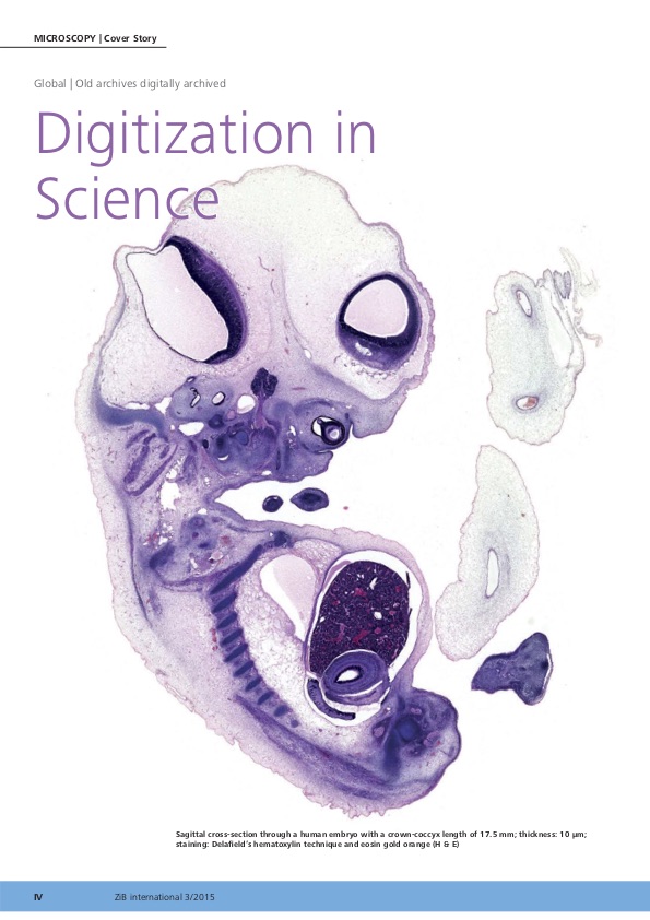
| 13:00, 13 April 2019 | EN Micro III-2015 Hill.jpg (file) |  |
109 KB | Z8600021 | 1 | |
| 11:31, 12 April 2019 | WNT3A transfer via filopodia.png (file) |  |
1.73 MB | Z8600021 | ==WNT3A transfer via filopodia== (a-c) Fluorescence photomicrographs of adjacent HeLa cells expressing RUSH-eGFP-WNT3A at indicated time points after biotin addition. From S7 video. (a) WNT3A is in the Golgi after biotin treatment. Inset showing WNT3A... | 1 |
| 10:55, 12 April 2019 | Acute otitis media infection age graph01.png (file) |  |
77 KB | Z8600021 | Fig 3. Age range of AOM infections. Each line represents an individual patient. N = 162 represents all patients who had at least one AOM infection. https://doi.org/10.1371/journal.pone.0212777.g003 acute otitis media (AOM) infection Copyright: © 201... | 1 |
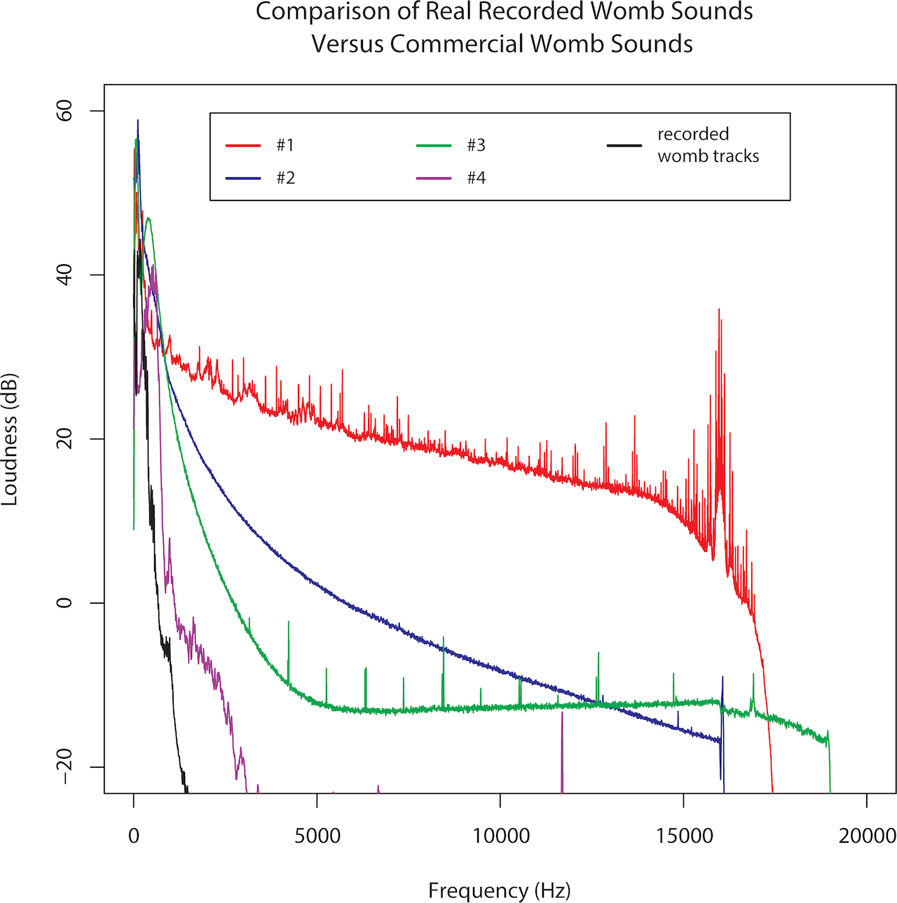
| 10:44, 12 April 2019 | Third trimester maternal womb sounds 01.mp3 (file) |  |
6.93 MB | Z8600021 | ==Maternal externally recorded womb sound recording== ===Reference=== {{#pmid:29746604}} ====Copyright==== © 2018 Parga et al. This is an open access article distributed under the terms of the Creative Commons Attribution License, which permits unr... | 1 |
| 10:40, 12 April 2019 | Third trimester sounds 01.png (file) |  |
335 KB | Z8600021 | 2 | |
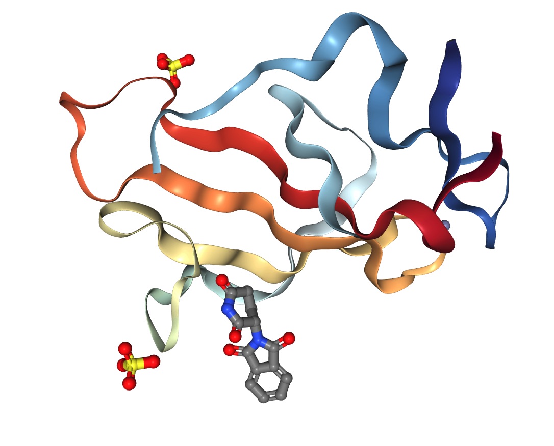
| 14:16, 11 April 2019 | Murine Cereblon in complex with Thalidomide.jpg (file) |  |
120 KB | Z8600021 | ==Crystal Structure of Murine Cereblon in Complex with Thalidomide== ===Reference=== Cite images created with the PDB ID and associated publication, NGL Viewer (AS Rose et al. (2018) NGL viewer: web-based molecular graphics for large complexes. Bioinf... | 1 |
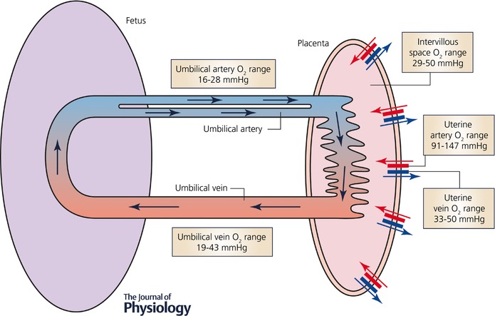
| 12:31, 10 April 2019 | Placental imaging 01.jpg (file) |  |
59 KB | Z8600021 | ==Placental Oxygen Levels== Simplified schematic diagram of the maternal and fetal placental circulations, showing the major compartments and published attributed in vivo oxygen values. :'''Links:''' {{placenta}} | {{trophoblast}} ===Reference===... | 1 |
| 12:18, 10 April 2019 | Placental imaging 03A.jpg (file) |  |
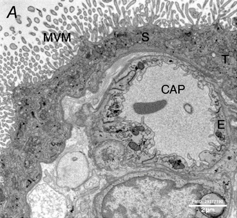
181 KB | Z8600021 | ==Placental Imaging== A transmission electron micrograph of terminal villi showing microvillous membrane (MVM), an underlying capillary (CAP), a syncytiotrophoblast (S), a trophoblast (T) and endothelium (E). :'''Links:''' {{placenta}} | [[:File:Pl... | 1 |
| 12:11, 10 April 2019 | Placental imaging 03.jpg (file) |  |
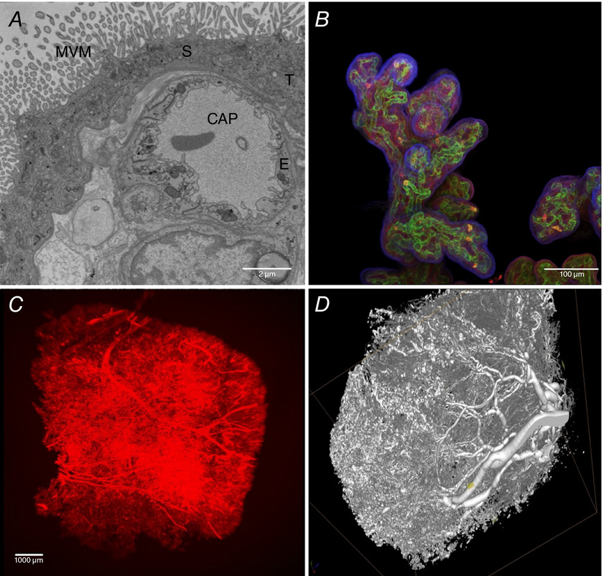
227 KB | Z8600021 | ==Placental Imaging== A, a transmission electron micrograph of terminal villi showing microvillous membrane (MVM), an underlying capillary (CAP), a syncytiotrophoblast (S), a trophoblast (T) and endothelium (E). B, projection of an imaged stack (whole... | 1 |
| 10:39, 9 April 2019 | Prentiss1906 fig07.jpg (file) |  |
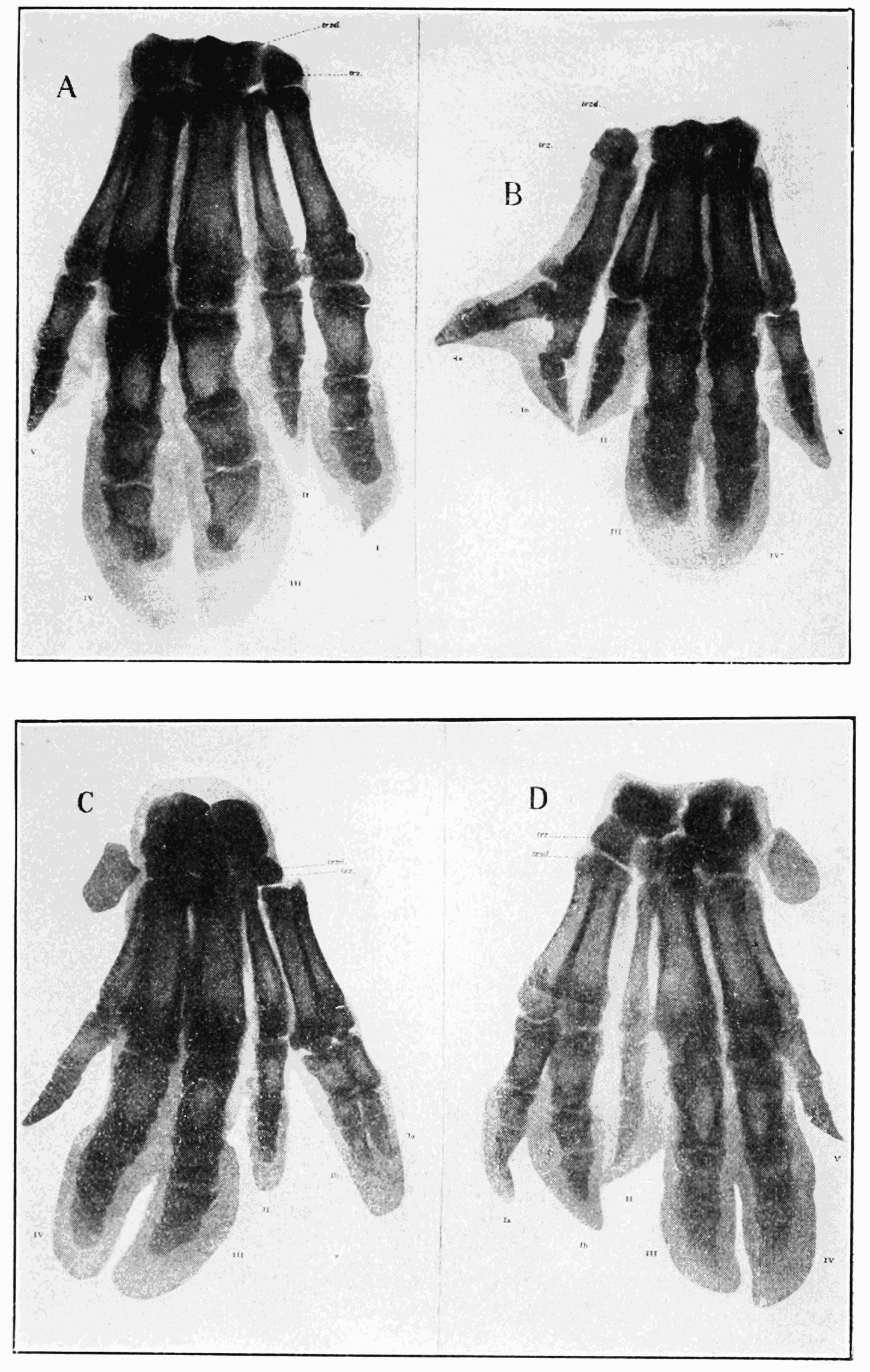
324 KB | Z8600021 | 1 | |
| 10:38, 9 April 2019 | Prentiss1906 fig06.jpg (file) |  |
138 KB | Z8600021 | 1 | |
| 10:37, 9 April 2019 | Prentiss1906 fig05.jpg (file) |  |
988 KB | Z8600021 | 1 | |
| 10:36, 9 April 2019 | Prentiss1906 fig04.jpg (file) |  |
490 KB | Z8600021 | 1 | |
| 10:35, 9 April 2019 | Prentiss1906 fig03.jpg (file) |  |
168 KB | Z8600021 | 1 | |
| 10:35, 9 April 2019 | Prentiss1906 fig02.jpg (file) |  |
159 KB | Z8600021 | 1 | |
| 10:34, 9 April 2019 | Prentiss1906 fig01.jpg (file) |  |
417 KB | Z8600021 | {{Ref-Prentiss1906}} | 1 |
| 21:28, 1 April 2019 | Wood-Jones1934 fig05.jpg (file) |  |
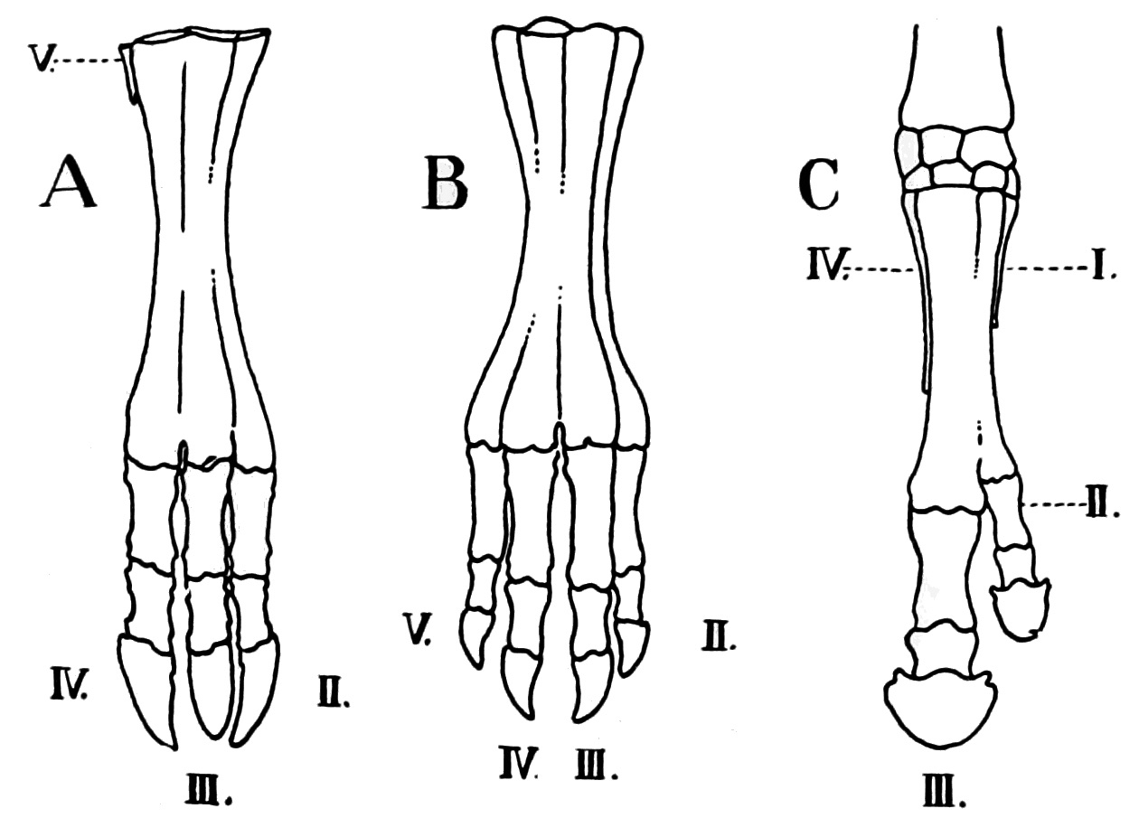
270 KB | Z8600021 | {{Ref-Wood-Jones1934}} | 1 |
| 21:27, 1 April 2019 | Wood-Jones1934 fig04.jpg (file) |  |
94 KB | Z8600021 | {{Ref-Wood-Jones1934}} | 1 |
| 21:24, 1 April 2019 | Wood-Jones1934 fig03.jpg (file) |  |
41 KB | Z8600021 | {{Ref-Wood-Jones1934}} | 1 |
| 21:23, 1 April 2019 | Wood-Jones1934 fig02.jpg (file) |  |
149 KB | Z8600021 | {{Ref-Wood-Jones1934}} | 1 |
| 21:20, 1 April 2019 | Wood-Jones1934 fig01.jpg (file) |  |
76 KB | Z8600021 | {{Ref-Wood-Jones1934}} | 1 |
| 14:16, 30 March 2019 | 2019 Foundations Lecture - Introduction to Human Development.pdf (file) |  |
4.31 MB | Z8600021 | Category:PDFCategory:MedicineCategory:2019 | 1 |
| 10:59, 29 March 2019 | Waldo Shumway.jpg (file) |  |
616 KB | Z8600021 | ==Waldo Shumway (1891-1956)== Born - 8 May 1891 New Brunswick, Middlesex County, New Jersey, USA Death - 8 Mar 1956 (aged 64) Manhattan, New York County (Manhattan), New York, USA {{Footer}} | 1 |
| 13:31, 27 March 2019 | Hair stem cells 01.jpg (file) |  |
123 KB | Z8600021 | ==Hair Stem Cells Niche== The hair follicle is a prominent example of how cell biology can help understand tissue homeostasis. Each hair follicle requires a niche of stem cells to undergo cyclical bouts of hair growth. The schematic (left) depicts t... | 1 |
| 16:50, 19 March 2019 | Trisomy 10 mosaicism cardiovascular abnormalities.jpg (file) |  |
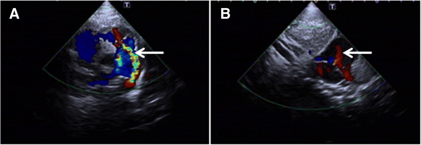
147 KB | Z8600021 | ==Trisomy 10 mosaicism Cardiovascular Abnormalities== Echocardiogram showing: a - a patent ductus arteriosus b - patent foramen ovale :'''Links:''' {{Trisomy mosaicism}} ===Reference=== {{#pmid:30081864}} ====Copyright==== Open Access This art... | 1 |
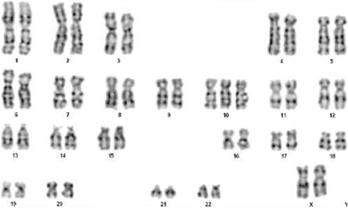
| 16:45, 19 March 2019 | Trisomy 10 mosaicism karyotype.jpg (file) |  |
76 KB | Z8600021 | Karyotype analysis: analysis of 100 split phase, 42 mitotic phase, karyotype of 47, XX ===Reference=== ====Copyright==== Open Access This article is distributed under the terms of the Creative Commons Attribution 4.0 International License (http://cr... | 1 |
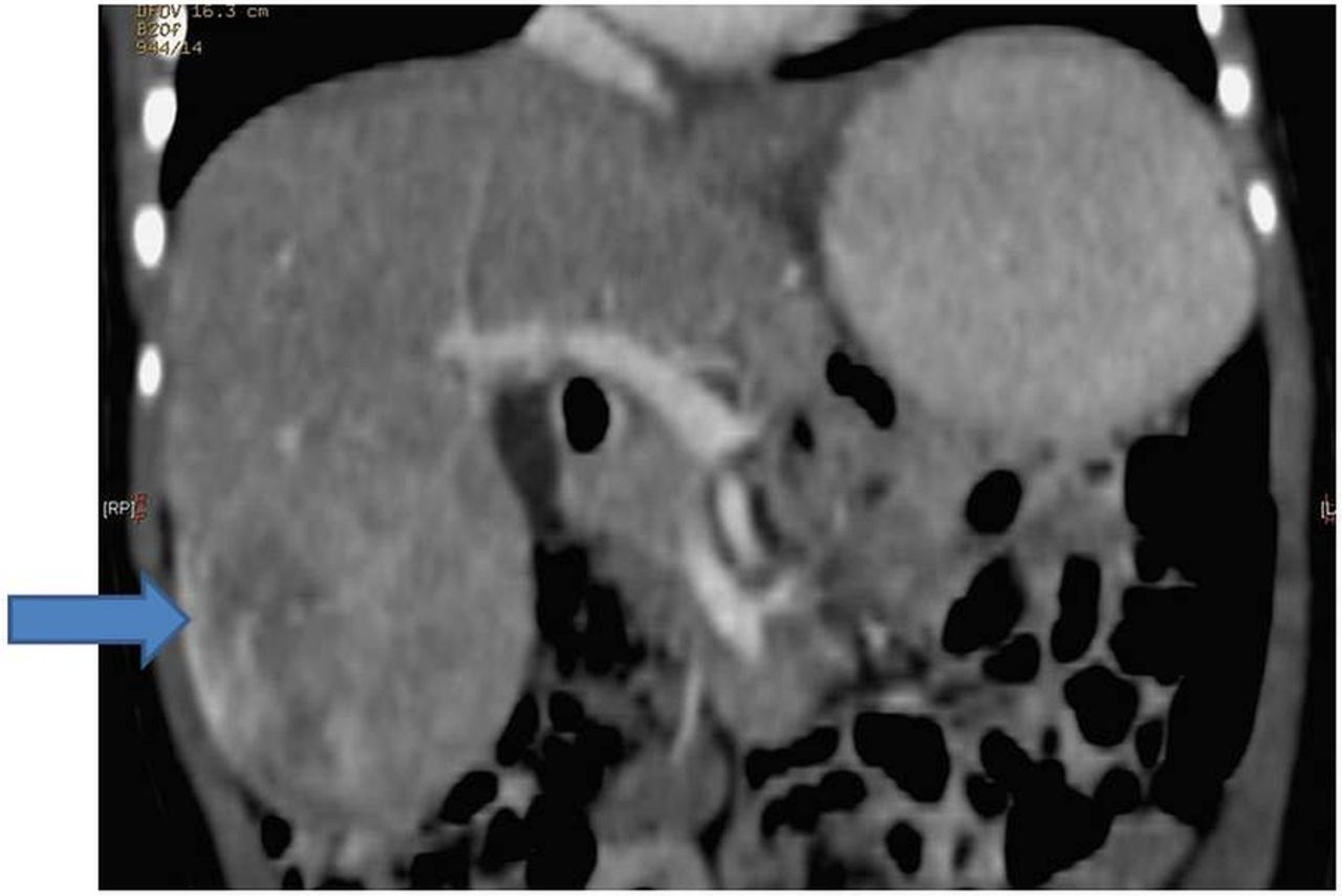
| 15:48, 19 March 2019 | Mosaic Trisomy 18 hepatoblastoma CT.jpg (file) |  |
79 KB | Z8600021 | ==Mosaic Trisomy 18 Hepatoblastoma== Diagnostic CT of the abdomen. {{#pmid:26795740}} Casereports-2016-January-2016---F2.large.jpg | 1 |
| 11:01, 19 March 2019 | Terminologia Embryologica User Guide.pdf (file) | |
269 KB | Z8600021 | Category:PDF | 1 |
| 16:15, 18 March 2019 | Angle1918 plate17.jpg (file) |  |
348 KB | Z8600021 | Angle1918 | 1 |
| 11:32, 15 March 2019 | Fetal week 12 head 14.jpg (file) |  |
95 KB | Z8600021 | 2 | |
| 11:31, 15 March 2019 | Fetal week 12 head 13.jpg (file) |  |
77 KB | Z8600021 | 2 | |
| 11:31, 15 March 2019 | Fetal week 12 head 12.jpg (file) |  |
47 KB | Z8600021 | 2 | |
| 10:50, 15 March 2019 | Fetal week 12 head 11.jpg (file) |  |
37 KB | Z8600021 | 1 | |
| 10:46, 15 March 2019 | Fetal week 12 head 1 icon.jpg (file) |  |
9 KB | Z8600021 | 1 | |
| 10:42, 15 March 2019 | Fetal week 12 head 01.jpg (file) |  |
48 KB | Z8600021 | 12wk-6605-lat-Trans-Diewert-UBC.avi | 1 |
| 10:07, 15 March 2019 | Fetal week 12 head 01.mp4 (file) | |
1,004 KB | Z8600021 | 12wk_6605_lat_Trans_Diewert_UBC.avi (398Wx398H) converted with handbrake 12wk_6605_lat_Trans_Diewert_UBC.mp4 | 1 |
| 11:14, 13 March 2019 | Catshark egg case stage 1.jpg (file) |  |
81 KB | Z8600021 | 1 | |
| 11:14, 13 March 2019 | Catshark egg case stage 2.jpg (file) |  |
58 KB | Z8600021 | 1 | |
| 11:13, 13 March 2019 | Catshark egg case stage 3.jpg (file) |  |
76 KB | Z8600021 | 1 | |
| 11:13, 13 March 2019 | Catshark egg case stage 4.jpg (file) |  |
98 KB | Z8600021 | 1 | |
| 11:13, 13 March 2019 | Catshark egg case stage 5.jpg (file) |  |
97 KB | Z8600021 | 1 | |
| 11:13, 13 March 2019 | Catshark egg case stage 6.jpg (file) |  |
106 KB | Z8600021 | 1 | |
| 11:12, 13 March 2019 | Catshark egg case stage 7.jpg (file) |  |
95 KB | Z8600021 | Fig 7. The inside of the S. stellaris egg case at stage 7. A: Position of placoid denticles and banded patterns on the body (dorsal view). B: Fully developed eye and greater pigmentation on skin (lateral view). C: The magnified head and fully consumed... | 1 |
| 11:03, 13 March 2019 | Catshark egg case stages1-7.jpg (file) |  |
94 KB | Z8600021 | catshark, Scyliorhinus stellaris ==Seven key developmental stages of catshark Scyliorhinus stellaris inside the egg case== Stage 1 was characterized by no visible embryo. Stage 2 was identified as an early embryo formed on the surface of the egg yolk... | 1 |