Search results
From Embryology
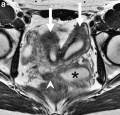
File:Female genital and ureter abnormality 01.jpg '''a''' Axial T2-W image demonstrates two widely separate uterine horns (large arrows), an obs ...age 2]] | [[:File:Female genital and ureter abnormality 03.jpg|Coronal T2-W image]] | [[Genital System - Abnormalities]] | [[Renal System - Abnormaliti(766 × 732 (86 KB)) - 08:25, 8 June 2017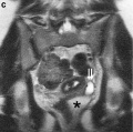
File:Female genital and ureter abnormality 03.jpg '''c''' Coronal T2-W image demonstrates the dilated left ureter (small arrows) inserting ectopic ...age 2]] | [[:File:Female genital and ureter abnormality 03.jpg|Coronal T2-W image]] | [[Genital System - Abnormalities]] | [[Renal System - Abnormaliti(766 × 762 (79 KB)) - 08:31, 8 June 2017
File:John W. Saunders Jr.jpg ==John W. Saunders Jr (1919 - 2015)==(400 × 587 (13 KB)) - 12:51, 7 September 2017
File:Female- OHVIRA syndrome 01.jpg '''a''' Axial T2-W image demonstrates two widely separate uterine horns (large arrows), an obs ...ht hemivagina (arrowhead), and a dilated left ureter (small arrows). On T1-W images, the fluid in the obstructed left hemivagina and the dilated left ur(340 × 1,000 (76 KB)) - 10:40, 16 July 2012
File:Mall1908a fig10.jpg X 8 times. L, liver; S, stomach; W, Wolffian body; SC, suprarenal body.(1,280 × 1,497 (344 KB)) - 12:12, 29 July 2018
File:Figure 1 – Immunohistochemical staining of β-catenin in spinal cord at different gestational ages.png “L, m” are magnifications with the microglial cells of “L, M”. E: Embryos; W: Weeks; M: Months; EP: Epithelial Ma, W., Yang, J. W., Gao, Y., Luo, T., Cheng, J. R., Wang, D. Y., ... & Li, L. Y. (2015). Expr(587 × 427 (691 KB)) - 12:30, 19 August 2016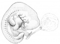
File:Minot1897 fig040.jpg After W. His.(1,236 × 905 (183 KB)) - 16:49, 27 July 2015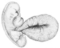
File:Minot1897 fig039.jpg After W. His.(1,020 × 816 (178 KB)) - 16:43, 27 July 2015
File:Figure1–Immunohistochemical staining of β-catenin in spinal cord at different gestational ages.png “L, m” are magnifications with the microglial cells of “L, M”. E: Embryos; W: Weeks; M: Months; EP: Epithelial Ma, W., Yang, J. W., Gao, Y., Luo, T., Cheng, J. R., Wang, D. Y., ... & Li, L. Y. (2015). Expr(587 × 427 (691 KB)) - 12:38, 19 August 2016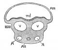
File:Minot1889 fig02.jpg modified from W. His. Am, amnion; md, medullary groove; v, v, veins; A, A, umbilical arteri(760 × 650 (91 KB)) - 11:51, 8 May 2018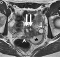
File:Female genital and ureter abnormality 02.jpg ...ht hemivagina (arrowhead), and a dilated left ureter (small arrows). On T1-W images, the fluid in the obstructed left hemivagina and the dilated left ur ...age 2]] | [[:File:Female genital and ureter abnormality 03.jpg|Coronal T2-W image]] | [[Genital System - Abnormalities]] | [[Renal System - Abnormaliti(766 × 733 (78 KB)) - 08:31, 8 June 2017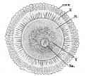
File:Minot1897 fig002.jpg ...radiata. Z, Zona pellucida. PI, Protoplasm. Y, Yolk. nu, Nucleus. — (After W. Nagel.)(828 × 746 (162 KB)) - 07:27, 5 April 2014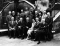
File:International Institute of Embryology, London 1938.jpg Top row (left to right): T. Thomson Flynn (Belfast); H. M. W. Woerdeman (Amsterdam); Hans Bluntschli (Bern); Jan Florian (Brno). Photograph sent by Fritz Strauss to Harland W. Mossman and is curated in Mossman's collection in Madison, Wisconsin.(1,000 × 763 (224 KB)) - 14:31, 4 July 2015
File:Bloxam1840-plate04.jpg Bloxam, W. On The Structure Of The Human Placenta, And Its Connexion With The Uterus.(1,438 × 1,470 (491 KB)) - 15:46, 24 September 2015
File:Gray1110 common male female genital.gif ...ary or testis is formed. ug. Sinus urogenitalis. W. Left Wolffian body. w, w. Right and left Wolffian ducts. ...rus. The uterine tube of the right side is marked m. v. Vulva. va. Vagina. W. Scattered remains of Wolffian tubes near it (paroöphoron of Waldeyer).(276 × 800 (55 KB)) - 18:13, 5 August 2009
File:Foster Balfour Sedgwick and Heap 1883.jpg Foster, M., Balfour, F. M., Sedgwick, A., & Heape, W. (1883). '''The Elements of Embryology.''' (2nd ed.). London: Macmillan and By Foster, M., Balfour, F. M., Sedgwick, A., & Heape, W. (1883) [[Book - The Elements of Embryology - Volume 1|The History of the C(716 × 1,057 (89 KB)) - 21:36, 28 March 2012
File:Rugh 045.jpg Courtesy, W. R. Duryee, 1950, Ann. N. Y. Acad. Sci., 50, Art. 8.(1,098 × 1,000 (244 KB)) - 09:23, 12 April 2013
File:Mall1891 Fig02.jpg * R W . - foramen of Winslow;(600 × 347 (45 KB)) - 16:29, 18 August 2015
File:Wen1928-Fig01.jpg ...yo H1093 (fig. 1, B) was obtained by Dr. Garrette Van Sweringen and Dr. W. W. Duemling, of Fort Wayne, Indiana. The clinical record gives the following ...X 16.6. Embryo H984 (fig. 1, C) was donated to the collection by Drs. E. W. Rawson and B. H. Foreman of Tacoma, Washington.(1,280 × 783 (80 KB)) - 23:17, 21 April 2016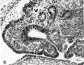
File:Faulconer1951 fig06.jpg ...ing to the dilated lumen, with structure suggesting :1 nephrostomal canal. W, Wolffian duct. X 300.(1,102 × 851 (253 KB)) - 14:50, 4 June 2016


