Search results
From Embryology
File:Mouse hematopoietic stem cell.gif ...ansitions from the fetal liver to the spleen and bone marrow late in fetal development. To date, experiments have not been performed to evaluate functionally the ...]] [[Category:Mouse]] [[Category:Stem Cell]] [[Category:Blood]] [[Category:Liver]][[Category:Bone]][[Category:Spleen]][[Category:Graph]](600 × 595 (40 KB)) - 11:27, 1 May 2019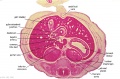
File:Stage 22 image 084.jpg [[Gastrointestinal Tract Development]] * Liver, Hepatic ducts. Gallbladder.(1,000 × 660 (136 KB)) - 14:04, 4 May 2013
File:Hepatitis B virus.jpg ...can be lifelong, known as cirrhosis (scarring) of the liver, liver cancer, liver failure, and death. Hepatitis B vaccine is available for all age groups to [[Category:Virus]] [[Category:Abnormal Development]] [[Category:Environmental Abnormalities]](700 × 1,030 (123 KB)) - 20:43, 7 November 2011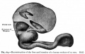
File:Bailey264.jpg ==Fig. 264. Reconstruction of the liver and intestine of a human embryo of 17 mm== ...definite parts of the small intestine. The first loop is the duodenum, the development of which has already been considered, and which maintains practically its o(881 × 562 (77 KB)) - 12:27, 15 April 2011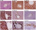
File:Histology of human embryonic liver at 11 weeks.png ==Histology of human embryonic liver at 11 weeks== Paraffin-embedded sections of human embryonic liver at 11 weeks (9 weeks of gestation) stained for Hematoxylin and Eosin (H&E),(1,988 × 1,642 (4.17 MB)) - 22:02, 8 November 2014
File:Gray0982b.jpg Sketches in profile of two stages in the development of the human digestive tube. (His.) A X 30. B X 20. ...in the formation of the diaphragm, while the caudal portion into which the liver grows forms the ventral mesogastrium.(427 × 393 (20 KB)) - 14:27, 28 April 2011
File:Gray0982a.jpg Sketches in profile of two stages in the development of the human digestive tube. ({{His}}) A X 30. B X 20. ...in the formation of the diaphragm, while the caudal portion into which the liver grows forms the ventral mesogastrium(427 × 393 (18 KB)) - 13:55, 13 August 2016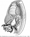
File:Bailey266.jpg * L - liver ...definite parts of the small intestine. The first loop is the duodenum, the development of which has already been considered, and which maintains practically its o(731 × 913 (178 KB)) - 16:31, 15 April 2014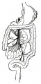
File:Gray0988.jpg ...arrested, so that in the adult it may be found lying immediately below the liver instead of in the right iliac region. ...] | [[:File:Gray0982b.jpg|Image - Late Week 4]] | [[Gastrointestinal Tract Development]] | [[Endoderm]](397 × 800 (48 KB)) - 15:10, 28 April 2011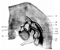
File:Hertwig1892 fig314.jpg neck measurement (embryo R, {{His}}), to elucidate the development of the pericardio-thoracic cavity and the diaphragm, after His. ...vein ; rj, jugular vein ; lg. lung ; z + I, fundament of the diaphragm and liver ; ilk, mandible.(983 × 828 (206 KB)) - 15:27, 21 February 2015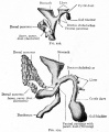
File:Bailey278 279.jpg ==Figs. 278 and 279. From models of the developing liver and pancreas of rabbit embryos== The Development of the Pancreas(669 × 812 (85 KB)) - 11:22, 15 April 2011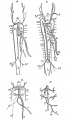
File:Quain594.jpg ==Fig. 594. Diagrams illustrating the development of the Great Veins== '''B''', veins of the liver at a somewhat earlier period.(592 × 1,000 (87 KB)) - 10:52, 8 June 2014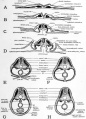
File:Patten054.jpg In considering the early development of the heart (Chapter IX) the formation of the dorsal and ventral mesocardi ...form the pericardial region of the body cavity (Figs. 24 and 55). Later in development the ventral mesentery farther caudally disappears so that caudally as well(764 × 1,060 (179 KB)) - 09:11, 29 July 2011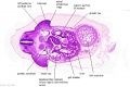
File:Stage 13 image 076.jpg [[Cardiovascular System Development]] [[Gastrointestinal Tract Development]](1,000 × 667 (120 KB)) - 13:12, 4 May 2013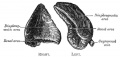
File:Image1184.jpg ...face is devoid of peritoneum, and is in relation with the bare area of the liver near its lower and medial angle, while its inferior portion is covered by p :'''Links:''' [[Endocrine - Adrenal Development|Adrenal Development]](800 × 378 (36 KB)) - 17:21, 25 March 2015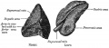
File:Image1183.jpg ...face is devoid of peritoneum, and is in relation with the bare area of the liver near its lower and medial angle, while its inferior portion is covered by p :'''Links:''' [[Endocrine - Adrenal Development|Adrenal Development]](800 × 351 (29 KB)) - 17:22, 25 March 2015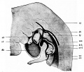
File:Hertwig1892 fig320.jpg ...surement ([[:File:Keibel Mall 034b.jpg|embryo R]], His), to illustrate the development of the pericardio-thoracic cavity and the diaphragm, after {{His}}. ...; vj, jugular vein ; Ig, lung ; z + I, fundament of the diaphragm and the liver ; uk, lower jaw.(970 × 824 (179 KB)) - 15:42, 21 February 2015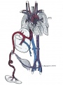
File:Gray0502.jpg :'''Links:''' [[Placenta Development]] | [[Cardiovascular System Development]] ...fore entering the inferior vena cava by the hepatic veins; some enters the liver directly, and is carried to the inferior cava by the hepatic veins; the rem(1,000 × 1,329 (215 KB)) - 21:34, 9 June 2014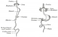
File:Gray0983.jpg Front view of two successive stages in the development of the digestive tube. (His.) (See enlarged image) ...in the formation of the diaphragm, while the caudal portion into which the liver grows forms the ventral mesogastrium.(800 × 517 (38 KB)) - 15:39, 28 April 2011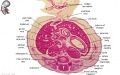
File:Embryo stage 22 F1L.jpg * gastrointestinal tract development ** associated organs - pancreas, liver(619 × 389 (79 KB)) - 11:33, 31 May 2010


