Search results
From Embryology
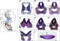
File:Mouse E10.5 Nav2 expression.jpg Nav2 is expressed in the mesencephalic tract, cranial and dorsal root ganglia and neural tube at E10.5. (A-F) Nav2 mRNA expressio ...D2, E2) overlaps with that of the Nav2 riboprobe in the ganglia of cranial nerve IX (D1) and X (E1).(1,200 × 818 (186 KB)) - 18:59, 14 April 2016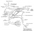
File:Gray0788.jpg (N. Facialis; Seventh Nerve; CN VII) ...edial, immediately to the lateral side of the sensory part is the acoustic nerve.(815 × 750 (91 KB)) - 00:21, 13 October 2015
File:Bailey361.jpg [[Category:Cranial Nerve]](815 × 662 (86 KB)) - 23:02, 4 September 2014
File:His1897 fig17.jpg ...the wall of the brain tube at the level of the origin of the fifth cranial nerve== ...wing to their having turned downwards to form the spinal root of the fifth nerve.(1,000 × 1,200 (303 KB)) - 15:25, 18 January 2016
File:His1897 plate03.jpg ...mbryo of four weeks. It shows the early condition of the roots of a spinal nerve. A diagrammatic representation of this is given in [[:File:His1897 fig02.jp ...the neuroblasts in the wall of the tube, growing out to form the anterior nerve-root.(1,738 × 2,808 (977 KB)) - 15:26, 18 January 2016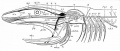
File:Bailey367.jpg ==Fig. 367. Diagram showing the principal branches of the cranial nerves in a fish== ...ial clefts; I, II, III, IV, VI, the first, second, third, fourth and sixth cranial nerves. The remaining nerves are differently shaded.(1,034 × 440 (101 KB)) - 08:19, 16 February 2016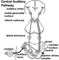
File:Auditory neural pathway.jpg ...ar nerve, acoustic nerve) part of the vestibulocochlear nerve (8th cranial nerve, CN VIII)(450 × 457 (46 KB)) - 18:33, 20 October 2013
File:Taste pathway.jpg ...s within the taste buds receive these chemical stimulus and convert into a nerve impulse. 3. this nerve impulse travels from the oral cavity cranial nerves towards the brain.(2,766 × 1,143 (783 KB)) - 00:19, 18 January 2013
File:Stage 22 image 162.jpg * The division of the VIII cranial nerve into a vestibular and auditory (spiral ganglia) component. * The eighth cranial nerve (VIII) exiting through the space known as the internal auditory meatus.(1,000 × 657 (176 KB)) - 12:00, 28 April 2011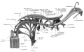
File:Keibel Mall 2 032.jpg ==Fig. 32. Reconstruction showing the cranial nerves in a 10 mm human embryo== ...similar but smaller tract is formed by the entering fibres of the acoustic nerve, which at this time consists mostly of vestibular fibres. In addition to th(1,200 × 750 (139 KB)) - 11:20, 8 October 2018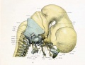
File:Lewis1920 fig08.jpg [[Category:Neural]][[Category:Cranial Nerve]](1,000 × 765 (81 KB)) - 10:55, 5 July 2014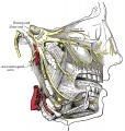
File:Gray0778.jpg ===Maxillary Nerve=== ...the lower eyelid, and the upper lip, joining with filaments of the facial nerve.(600 × 630 (101 KB)) - 10:54, 25 June 2018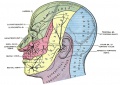
File:Gray0784.jpg ...bution of the three divisions of the fifth nerve (N. Trigeminus, Trifacial Nerve, '''{{CN V}})'''. ...l nerve and is the great sensory nerve of the head and face, and the motor nerve of the muscles of mastication. It emerges from the side of the pons, near i(851 × 600 (137 KB)) - 10:57, 25 June 2018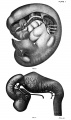
File:Mall1905 plate01-2.jpg ...reconstructed; the tenth and twelfth cranial nerves and the flrst cervical nerve by Dr. Streeter. ...he circle of Willis throughout that of the fore-brain. The position of the cranial nerves and otic vesicle is given in Fig. 9.(1,187 × 2,000 (266 KB)) - 10:44, 15 January 2017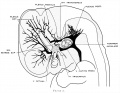
File:Streeter1921 fig02.jpg ...anial (primary head-vein). Median to the second division of the trigeminal nerve can he seen the plexiform maxillary vein, which drain the structure of the [[Category:Hearing]][[Category:Cranial Nerve]](1,286 × 1,000 (138 KB)) - 10:27, 16 May 2017
File:Human Stage14 neural02.jpg {{Cranial Nerve Table}}(1,375 × 2,048 (506 KB)) - 17:55, 14 April 2016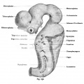
File:Kollmann636.jpg ==The exits of the cranial nerves in the brain of a human embryo of 10.2 mm CRL== ...auditory nerve. The type of the spinal nerves still follows the oculomotor nerve with respect to the exit point.(759 × 756 (65 KB)) - 13:53, 29 January 2016
File:Stage 22 image 218.jpg ...- cochlear component of the ganglia (vestibulocochlear nerve, 8th cranial nerve, CN VIII) top of the image.(1,200 × 730 (308 KB)) - 11:44, 14 June 2016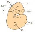
File:Day 11 Closure of lens vesicle.JPG ...esicle, G7 - G8= ganglion of the 7th and 8th cranial nerve, G5= trigeminal nerve ganglion, 1= 1st branchial bar, 2= 2nd branchial bar(560 × 528 (27 KB)) - 13:19, 18 August 2014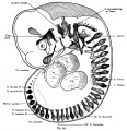
File:Kollmann647.jpg the eye, the middle cranial beams and the maze bubbles L;: tonic Xll means the hypoglossal nerve;(897 × 926 (188 KB)) - 13:17, 29 January 2016


