Search results
From Embryology
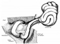
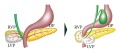
File:Frazer1915 fig10.jpg ...e and below the cut vitelline vein. The mesoduodenum is visible behind the duodenum and below the foramen of Winslow. Cf. fig. 8.(1,000 × 733 (155 KB)) - 05:42, 9 January 2017
File:Jejunum and ileum cartoon.jpg ...egions starting from the stomach they are the [[:File:Duodenum cartoon.jpg|duodenum]], [[:File:Jejunum and ileum cartoon.jpg|jejunum and ileum]]. | Duodenum (25 cm)(500 × 704 (45 KB)) - 12:04, 11 April 2019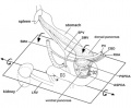
File:Human fetal pancreas anatomy cartoon.jpg Fetal topographical anatomy of the pancreatic head and duodenum with special reference to courses of the pancreaticoduodenal arteries. ...hesize a rotation along a left-right axis through the third portion of the duodenum in the later stage of development. In addition, the fourth portion is conne(455 × 376 (93 KB)) - 17:38, 6 April 2018- upper {{duodenum}} | valign=top|lower {{duodenum}}822 bytes (93 words) - 13:00, 17 April 2019
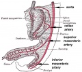
File:GIT blood supply.jpg # '''Foregut''' - celiac artery (Adult: pharynx, esophagus, stomach, upper duodenum, respiratory tract, liver, gallbladder pancreas) # '''Midgut''' - superior mesenteric artery (Adult: lower duodenum, jejunum, ileum, cecum, appendix, ascending colon, half transverse colon)(568 × 500 (47 KB)) - 12:48, 17 April 2019- Frazer JE. Note on Dr Hunter's Paper on Development of the Duodenum. (1927) {{J. Anat.}} 61: 356-9. [https://www.ncbi.nlm.nih.gov/pubmed/171041160 bytes (23 words) - 09:40, 4 February 2020
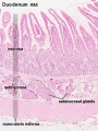
File:Intestine histology 005.jpg ==Duodenum==(400 × 533 (78 KB)) - 13:17, 3 April 2013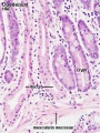
File:Intestine histology 007.jpg ==Duodenum==(400 × 533 (82 KB)) - 13:34, 3 April 2013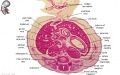
File:Embryo stage 22 F1L.jpg ** [[S#stomach|stomach]], [[D#duodenum|duodenum]], [[J#jejunum|jejunum]](619 × 389 (79 KB)) - 11:33, 31 May 2010- CONGENITAL CONSTRICTION OF THE DUODENUM DUE TO AN ABNORMAL FOLD OF THE ANTERIOR MESOGASTRIUM Reginald H. Jackson An168 bytes (22 words) - 20:33, 10 August 2020
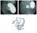
File:Duodenal atresia 02.jpg ('''a''') Upper gastrointestinal series showing a complete obstruction of the duodenum and contrast filling of anomalous bifurcated bile ducts (arrows). The small ...estinal series showing a complete obstruction at the second portion of the duodenum, and contrast was seen in the proximal jejunum which is located in the righ(765 × 682 (68 KB)) - 16:21, 16 April 2019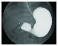
File:Duodenal atresia 01.jpg ...complete obstruction to the flow of contrast at the second portion of the duodenum. There is also contrast filling of the biliary tree above the duodenal bulb ...ructural developmental anomalies of duodenum] - ''Any congenital defect of duodenum that results from interference with the normal growth and differentiation o(750 × 592 (56 KB)) - 16:21, 16 April 2019- ...e of the greater part of the small and large intestines, continuity of the duodenum with the colon, absence of the left testis, epididymis and cord, and enormo743 bytes (102 words) - 10:16, 13 October 2020
- ...Gastrointestinal Tract]][[Category:Trachea]][[Category:Stomach]][[Category:Duodenum]]</noinclude>216 bytes (21 words) - 14:52, 23 January 2019
File:Annular pancreas.jpg ...rsists, and the right ventral pancreatic anlage does not rotate around the duodenum. The two ventral anlagen encircle the duodenum.(600 × 265 (16 KB)) - 17:36, 23 May 2016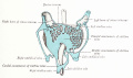
File:Arey1924 fig194.jpg ...ion in the liver, ventral to the duodenum; (2) a middle one, dorsal to the duodenum; and (3) a caudal one, ventral to it. There are thus formed about the gut a(1,200 × 710 (122 KB)) - 07:10, 24 October 2016- * Small intestine - including the duodenum distal to the opening of the bile duct182 bytes (30 words) - 12:55, 23 January 2019
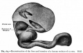
File:Bailey264.jpg ...al vein; 1 -6, primary bends in the long intestinal loop; 1 represents the duodenum. ...ned to become definite parts of the small intestine. The first loop is the duodenum, the development of which has already been considered, and which maintains(881 × 562 (77 KB)) - 12:27, 15 April 2011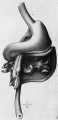
File:Bardeen1914-fig07.jpg Ventral view of a reconstruction of the stomach, duodenum, eaeeum, colon, rectum, bursa omentalis and rnesentery of fetus 40 mm long(451 × 936 (63 KB)) - 09:53, 3 October 2017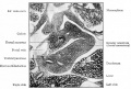
File:Bailey280.jpg ==Fig. 280. From a transverse section through the region of the duodenum of a pig embryo of 14 mm==(866 × 581 (133 KB)) - 18:14, 24 January 2011


