Uncategorised files
From Embryology
Showing below up to 50 results in range #1,251 to #1,300.
 Opac figure 10.jpg 505 × 243; 18 KB
 Opac figure 7.jpg 505 × 243; 18 KB
 Openoffice-export.jpg 569 × 243; 46 KB
 Openoffice-menu.jpg 378 × 459; 50 KB
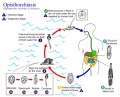 Opisthorchiasis.jpg 739 × 598; 84 KB
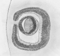 Optic cup at carnegie stage 17 .jpg 640 × 597; 297 KB
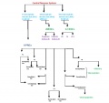 Origin of the craniofacial skeleton-1.jpg 585 × 549; 123 KB
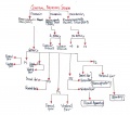 Origin of the craniofacial skeleton.jpg 2,304 × 2,039; 319 KB
 Orlando Charnock Bradley.jpg 563 × 600; 110 KB
 Osteocyte 01.jpg 269 × 233; 4 KB
 Osteocyte 02.jpg 524 × 403; 16 KB
 Outflow Tract A.ppt ; 453 KB
- Outflow Tract advanced draft.ppt ; 446 KB
- Outflow Tract intermediate draft.ppt ; 380 KB
 Ovary corpus luteum 01.jpg 1,028 × 576; 206 KB
 Ovary corpus luteum 02.jpg 1,028 × 609; 122 KB
 Ovine fetal artery hypoxia genes 01.jpg 1,129 × 1,000; 98 KB
 Oxidative Stress Response in Friedreich Ataxia.jpg 561 × 695; 178 KB
 PCP pathway leading to gastrulation.jpg 1,523 × 2,048; 128 KB
 PCP pathway leading to gastrulation during embryonic development.JPG 1,523 × 2,048; 128 KB
 PCP pathway leading to gastrulation in embryonic development.jpg 1,523 × 2,048; 128 KB
 PDA arterial photoplethysmography.jpg 600 × 358; 31 KB
 PMID--2518447journal.pone.0104728.g001A.jpg 1,206 × 908; 168 KB
 PRACTICAL CLASS 3 PROGRAM.pdf ; 131 KB
- PRACTICAL CLASS 7 PROGRAM.pdf ; 114 KB
- PRACTICAL CLASS 7 PROGRAM final.pdf ; 114 KB
 Padget1957 fig01.jpg 2,199 × 2,422; 821 KB
 Paeonia suffruticosa.jpg 640 × 480; 75 KB
 Palate.gif 550 × 400; 135 KB
 Palate 001 icon.jpg 192 × 190; 17 KB
 Palate 002 icon.jpg 192 × 190; 17 KB
 Papillae.jpg 900 × 600; 197 KB
 Paraben.png 629 × 1,100; 20 KB
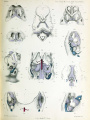 Parker1874 plate29.jpg 1,028 × 1,375; 171 KB
- Part 1 muscle development 2012.pdf ; 1.11 MB
- Part 2 Muscle Development 2012.pdf ; 552 KB
 Parvovirus infection childhood hands.jpg 700 × 460; 38 KB
 Paterson1900 plate3.jpg 1,280 × 1,085; 403 KB
 Pathophyis 1.jpg 588 × 637; 16 KB
 Pathophysiology & development.jpg 601 × 637; 15 KB
 Pathophysiology 2.jpg 588 × 637; 17 KB
 Patterns of kmrl mRNA in mouse embryo.jpg 480 × 513; 57 KB
 PdP.gif 2,880 × 1,800; 5.65 MB
 Pedigree chart.jpg 600 × 368; 44 KB
 Pedigree of Friedreich's Ataxia.PNG 618 × 384; 46 KB
 Pendred Syndrome Mondini Defect.jpg 1,800 × 590; 193 KB
 Pendred Syndrome Showing Mondini Defect.jpg 640 × 657; 56 KB
 Permalink Help 1. Open the Virtual Slide.jpg 800 × 658; 123 KB
 Permalink Help 2. Zoom to the Region of Interest.jpg 800 × 658; 85 KB


