Unused files
From Embryology
The following files exist but are not embedded in any page. Please note that other web sites may link to a file with a direct URL, and so may still be listed here despite being in active use.
Showing below up to 50 results in range #1,201 to #1,250.
 Hill1931 plate33.jpg 1,551 × 1,894; 731 KB
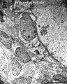 Human ovary follicle basement membrane EM01.jpg 558 × 697; 100 KB
 Fetal cardiac ultrasound 01.jpg 900 × 600; 88 KB
 Stage11 sem3a.gif 749 × 800; 371 KB
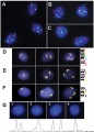 Human fetal neural aneuploidy.jpg 1,000 × 1,400; 134 KB
 Minot1897-463.jpg 354 × 312; 28 KB
 Minot1897-460.jpg 1,038 × 896; 113 KB
 Minot1897-459.jpg 975 × 460; 82 KB
 Minot1897-458.jpg 602 × 982; 59 KB
 Minot1897-462.jpg 431 × 551; 122 KB
 Minot1897-461.jpg 511 × 415; 63 KB
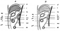 Minot1897-457.jpg 983 × 517; 92 KB
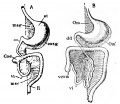 Minot1897-456.jpg 540 × 470; 52 KB
 Minot1897-455.jpg 533 × 613; 70 KB
 Minot1897-005.jpg 1,154 × 720; 166 KB
 RBC with hereditary persistence of fetal haemoglobin SEM01.jpg 1,200 × 816; 220 KB
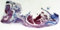 B050966-01.jpg 2,000 × 992; 681 KB
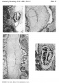 Odgers1937 plate02.jpg 1,430 × 2,000; 961 KB
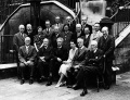 International Institute of Embryology, London 1938.jpg 1,000 × 763; 224 KB
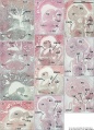 Heart human embryo CRL10mm 01.jpg 1,000 × 1,389; 537 KB
 The process of neurulation.mp4 ; 29.18 MB
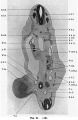 Barniville1914 fig21.jpg 1,023 × 1,569; 301 KB
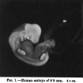 Barniville1914 fig01.jpg 765 × 758; 59 KB
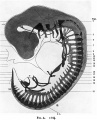 Barniville1914 figA.jpg 1,000 × 1,237; 269 KB
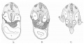 Thompson1908 02.jpg 1,444 × 753; 192 KB
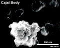 Cajal body EM.jpg 600 × 487; 29 KB
 Wyburn1937 table01.jpg 1,448 × 2,142; 548 KB
 Abnormal formation of fertilisation cones .jpg 256 × 256; 5 KB
- Cardiac Tutorial statistics.xls ; 33 KB
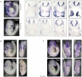 Mouse E9.5 neural crest beta actin KO.jpg 2,722 × 2,573; 732 KB
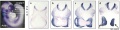 Mouse E9.5 neural crest - Crabp1.jpg 2,489 × 626; 217 KB
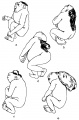 Nanagas1925-fig01.jpg 1,319 × 2,000; 251 KB
 Streeter1917-fig02.jpg 1,000 × 847; 276 KB
 Streeter1917-fig08-09.jpg 1,200 × 853; 218 KB
 Streeter1917-fig06-07.jpg 1,337 × 1,671; 639 KB
 Streeter1917-fig04-05.jpg 1,000 × 544; 124 KB
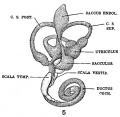 Streeter1917-fig05.jpg 556 × 544; 64 KB
 B2020649-170-2.jpg 929 × 1,506; 142 KB
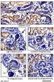 Mouse nephron stages 01.jpg 601 × 916; 217 KB
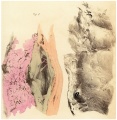 Bloxam1840-plate04.jpg 1,438 × 1,470; 491 KB
 DNA-biological target of radiation.jpg 542 × 245; 38 KB
 Normal ovary compared to polycystic ovary.png 2,636 × 948; 1.36 MB
 Method 1 maternal spindle transfer.png 1,200 × 804; 537 KB
 Method 2 pronuclear transfer.png 1,200 × 752; 592 KB
 Components and structure of Spermatozoa.jpeg 600 × 783; 67 KB
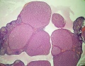 Rat ovary histology 01.jpg 1,200 × 938; 274 KB
 What is Oncofertility?.png 981 × 785; 187 KB
 Summary of Oncofertility.pdf ; 160 KB
 Woman's Uterus.gif 300 × 233; 26 KB


