Unused files
From Embryology
The following files exist but are not embedded in any page. Please note that other web sites may link to a file with a direct URL, and so may still be listed here despite being in active use.
Showing below up to 50 results in range #1,051 to #1,100.
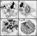 Fetal white blood cell.jpeg 792 × 757; 82 KB
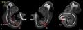 Mouse embryo E11 HNF3beta notochord marker 01.jpg 2,245 × 829; 129 KB
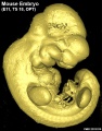 Mouse embryo E11 tomography 01.jpg 628 × 800; 82 KB
 Effect of DMSO on asymmetric cell division in mouse oocytes.jpg 600 × 1,079; 126 KB
 Mouse blastocyst.png 819 × 420; 58 KB
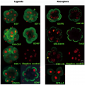 Antibody immunostaining of ligands and receptors in triploid human embryos.png 3,439 × 3,444; 7.13 MB
 Secondary Oocyte during in-vitro fertilisation.jpeg 800 × 558; 48 KB
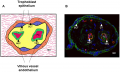 Trophoblast and villous epithelium of the placenta.png 2,058 × 1,241; 2.61 MB
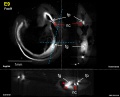 Mouse E9 Foxf1.jpg 1,420 × 1,143; 142 KB
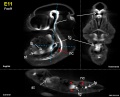 Mouse E11 Foxf1.jpg 1,420 × 1,143; 162 KB
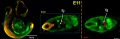 Mouse E11 Sox2 and Nkx2.1.jpg 2,229 × 720; 176 KB
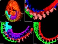 Mouse E9 cervical intersomitic vessels.jpg 2,989 × 2,300; 777 KB
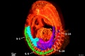 Mouse E9 cervical intersomitic vessels A.jpg 1,714 × 1,142; 209 KB
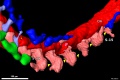 Mouse E9 cervical intersomitic vessels B.jpg 1,714 × 1,142; 175 KB
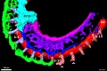 Mouse E9 cervical intersomitic vessels C.jpg 1,714 × 1,142; 245 KB
 Mouse E9 cervical intersomitic vessels D.jpg 1,714 × 1,142; 147 KB
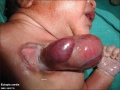 Ectopia cordis.jpg 800 × 603; 37 KB
 Sirenomelia 02.jpg 498 × 598; 14 KB
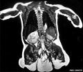 Caudal duplication syndrome.jpg 700 × 599; 47 KB
 Nail patella syndrome 02.jpg 809 × 525; 51 KB
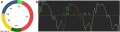 Fetal cardiac state diagram 01.jpg 1,200 × 321; 29 KB
 Fetal cardiac state diagram 02.jpg 1,200 × 458; 27 KB
 Fetal ultrasound ductal arch 01.jpg 800 × 533; 27 KB
 Fetal heart atrioventricular plane displacement 01.jpg 915 × 733; 55 KB
 Ovine fetal artery hypoxia genes 01.jpg 1,129 × 1,000; 98 KB
 Dmitry Mendelejew 1834-1907.jpg 847 × 1,200; 148 KB
 Mouse eye neural crest cornea 02.jpg 800 × 655; 121 KB
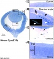 Mouse eye E18.jpg 884 × 977; 169 KB
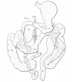 Roux1911 fig02.jpg 1,229 × 1,400; 175 KB
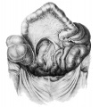 Roux1911 fig01.jpg 1,108 × 1,200; 264 KB
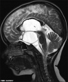 Rathke cleft cyst 01.jpg 493 × 600; 46 KB
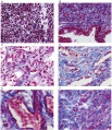 Fetal corpus cavernosum and corpus spongiosum 01.jpg 1,795 × 2,082; 919 KB
 PMID--2518447journal.pone.0104728.g001A.jpg 1,206 × 908; 168 KB
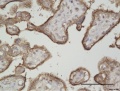 Human placenta vasohibin 2 expression.jpg 1,198 × 904; 165 KB
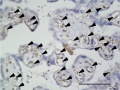 Human placenta vasohibin 1 expression.jpg 1,206 × 908; 168 KB
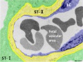 Mouse placenta blood vessel EM01.jpg 1,000 × 739; 132 KB
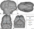 Mouse head E11.5 microCT 01.jpg 1,409 × 1,200; 308 KB
 Septation of the heart.jpg 2,258 × 705; 288 KB
 Early fetal valve development.jpg 1,000 × 612; 164 KB
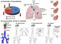 Lung Models Normal vs. Diseased.png 600 × 434; 211 KB
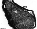 Human spermatozoa nucleus EM02.jpg 597 × 476; 52 KB
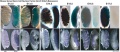 Mouse gonad Gcnf expression 01.jpg 1,947 × 843; 304 KB
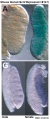 Mouse gonad Gcnf expression E12.5.jpg 331 × 785; 68 KB
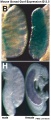 Mouse gonad Gcnf expression E13.5.jpg 332 × 784; 60 KB
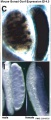 Mouse gonad Gcnf expression E14.5.jpg 334 × 784; 60 KB
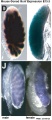 Mouse gonad Gcnf expression E15.5.jpg 338 × 782; 53 KB