Search results
From Embryology
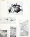
File:Mall Meyer1921 plate15.jpg ...nded by clot. Tr, trophoblast; L, leucocytes; Ch, chorion; V, villus; Coe, coelom. X50.(979 × 1,200 (182 KB)) - 09:32, 6 December 2012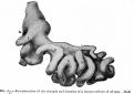
File:Bailey265.jpg ...e and form secondary loops, all of which push their way into the umbilical coelom where they remain until the embryo reaches a length of 40 mm. (compare [[Bo(906 × 644 (80 KB)) - 12:40, 15 April 2011
File:HHstage7.jpg | 1-3 somites; coelom(671 × 980 (72 KB)) - 12:35, 2 June 2017
File:Stage13 bf4.jpg * Larger cavity is chorionic space (coelom).(1,200 × 800 (132 KB)) - 16:38, 11 September 2014
File:Stage14 bf18.jpg * Larger cavity is chorionic space (coelom).(1,800 × 2,250 (549 KB)) - 17:28, 11 September 2014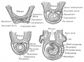
File:Rugh 107.jpg ...the two sides grows together ventrally to fuse below the foregut. Both the coelom and the continuous and related pericardial cavity arise as bilateral caviti(1,073 × 800 (188 KB)) - 10:35, 17 April 2013
File:Ingalls1920FigureA.jpg ...with the foregut; farther forward, in the median line, is the pericardial coelom with its contained heart.(1,000 × 504 (66 KB)) - 09:02, 3 February 2012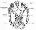
File:Keibel Mall 2 530.jpg ...he right side, is fused with the lateral plate and receives a cleft of the coelom. The rete peri-intestinale is well developed, since we are here in the regi(1,280 × 1,080 (239 KB)) - 17:15, 14 November 2018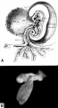
File:Hertig1946b fig09.jpg ...he communication between the exocoelomic space around the yolk-sac and the coelom (body cavity) within the embryo. Carnegie {{CE6488}}, sequence 2, X15. 104(800 × 1,481 (169 KB)) - 17:34, 7 August 2017
File:Odgers1937 plate02.jpg ...he yolk sac surrounded on either side and ventrally by the extra-embryonic coelom.(1,430 × 2,000 (961 KB)) - 21:52, 29 June 2015
File:Bremer1914 plate04.jpg ...erm. at the bottom of the drawing, znd the stumps of two villi (zd.).C'oe.,coelom;a, unlined space;b, angioblast cord. X circa 800.(643 × 1,000 (127 KB)) - 11:25, 29 July 2019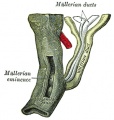
File:Gray1109.jpg ...responding Wolffian duct as a tubular invagination of the cells lining the coelom(464 × 487 (56 KB)) - 11:07, 22 November 2018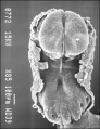
File:Stage 9 SEM1.jpg The intra-embryonic coelom develops in the middle of the lateral plate mesoderm. Note amniotic ectoder(347 × 450 (42 KB)) - 17:15, 30 July 2011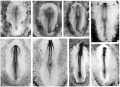
File:HHstage5-10.jpg | 1-3 somites; coelom(2,864 × 2,075 (734 KB)) - 16:26, 2 June 2017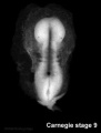
File:Stage9sm.jpg The intra-embryonic coelom develops in the middle of the lateral plate mesoderm. Note amniotic ectoder(287 × 378 (6 KB)) - 10:43, 9 August 2009
File:Stage9 ventral.jpg The intra-embryonic coelom develops in the middle of the lateral plate mesoderm. Note amniotic ectoder(291 × 384 (5 KB)) - 07:23, 7 April 2012
File:Stage9 dorsal.jpg The intra-embryonic coelom develops in the middle of the lateral plate mesoderm. Note amniotic ectoder(287 × 378 (5 KB)) - 07:26, 7 April 2012
File:Shaw1932 fig01.jpg Figs. 3 and 4. A, cerebral vesicle; B, heart; 0, coelom; D, intestine; E, primitive kidney; F, limb bud; G, dorsal aorta; H, neural(900 × 715 (59 KB)) - 17:08, 15 November 2017
File:Shaw1932 plate01.jpg Figs. 3 and 4. A, cerebral vesicle; B, heart; 0, coelom; D, intestine; E, primitive kidney; F, limb bud; G, dorsal aorta; H, neural(2,178 × 2,950 (454 KB)) - 17:07, 15 November 2017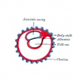
File:Gray0025.jpg ...is becomes expanded into a vesicle which projects into the extra-embryonic coelom.(500 × 500 (29 KB)) - 12:10, 25 February 2014


