Search results
From Embryology
Page title matches
File:Liver development signaling.jpg ==Overview of Mouse Liver Embryogenesis== :'''Links:''' {{liver}}(600 × 467 (45 KB)) - 16:46, 30 March 2019

Page text matches
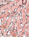
File:Liver-reticular fibre.jpg ==Liver Histology== Liver - Silver stain (reticulin)(700 × 875 (77 KB)) - 13:15, 9 March 2018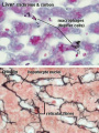
File:Liver- Kupffer cell and reticular fibre.jpg ==Liver Histology== Liver, rabbit - trichrome & carbon and Liver - reticulin(600 × 800 (49 KB)) - 13:08, 9 March 2018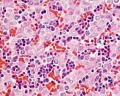
File:Histology-fetal liver HEx40.jpg ==Fetal Liver== This histology section of the fetal liver shows the prresence of large numbers of red blood cells (erythrocytes) and(1,000 × 800 (281 KB)) - 13:53, 23 February 2013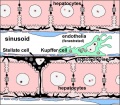
File:Liver-sinusoid colour cartoon.jpg ==Architecture of the Liver Sinusoid== ...endothelia and interspersed Kupffer cells, the resident macrophages of the liver.(600 × 523 (64 KB)) - 07:45, 4 November 2015
File:Thompson1908 02.jpg B, at level of liver bud; V., ventricle; 1>.C., pericanlial cavity; 8., stomach; H.B., liver bud; V.V., vltelline vein;(1,444 × 753 (192 KB)) - 22:57, 11 August 2015
File:Stage 22 image 083.jpg {{Virtual Slide Features - Stage 22 Liver}} [[Gastrointestinal Tract Development]](1,000 × 648 (125 KB)) - 18:42, 23 September 2015
File:Human liver week 9.jpg ==Human Fetal Liver== Paraffin-embedded sections of human embryonic liver at 9 weeks ({{GA}} 11 weeks.)(1,200 × 991 (425 KB)) - 13:19, 6 June 2014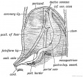
File:Keith1902 fig213a.jpg ==Fig. 213. The origin of the Peritoneal Ligaments connected with the Liver== ...esentery. Out of the ventral mesentery are formed all the ligaments of the liver (Fig. 213 A). These are the following:(854 × 800 (166 KB)) - 12:25, 22 July 2018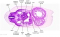
File:Stage 13 image 073.jpg * Coelomic canals change in outline, due to presence of liver. Lesser sac. * Liver embedded in septum transversum (ventral border of septum transversum contri(1,000 × 619 (118 KB)) - 07:37, 15 May 2014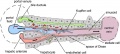
File:Liver plasmodium infection cartoon.jpg ==Model of Plasmodium Sporozoite Infection of the Mammalian Liver== ...ches of the portal vein and the hepatic artery, merges upon entry into the liver lobule at the portal field. The blood flows along the sinusoid and exits at(1,000 × 450 (78 KB)) - 10:33, 27 January 2019
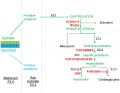
File:Zorn2008 fig01.jpg ==Fig, 1 Liver cell lineage== The cell lineage steps during hepatic development (red) from uncommitted endoderm to functional adult hepatocytes and biliary(1,200 × 1,158 (110 KB)) - 16:21, 20 August 2017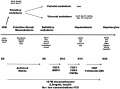
File:Liver hepatocyte from stem cell.png Overview of liver embryogenesis on which liver differentiation protocol is based. Genes specifically expressed at different steps during liver development are shown, in italics, under each step of lineage commitment. Cytokines use(600 × 444 (96 KB)) - 17:56, 25 August 2010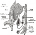
File:Gray0989.jpg ...he stomach and liver, and the falciform and coronary ligaments between the liver and the abdominal wall and diaphragm (Fig. 989). ...] | [[:File:Gray0982b.jpg|Image - Late Week 4]] | [[Gastrointestinal Tract Development]] | [[Endoderm]](700 × 685 (107 KB)) - 15:21, 28 April 2011
File:Bookshelf NBK27068.pdf This is the PDF version of the online StemBook chapter on liver development. Note that the information is specific to the date of creation (2008) of th Zorn AM. [https://www.ncbi.nlm.nih.gov/books/NBK27068/ Liver development]. 2008 Oct 31. In: StemBook [Internet]. Cambridge (MA): Harvard Stem Cell I(5.64 MB) - 13:05, 14 March 2017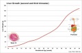
File:Fetal liver weight growth graph.jpg ==Human Fetal Liver Weight Growth== :{{Fetal Graphs}} | [[Gastrointestinal Tract - Liver Development|Liver Development]](800 × 521 (34 KB)) - 17:00, 23 April 2012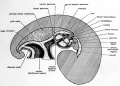
File:Patten055.jpg ...form the pericardial region of the body cavity (Figs. 24 and 55). Later in development the ventral mesentery farther caudally disappears so that caudally as well ...ver persists as the gastro-hepatic omentum, and the portion ventral to the liver persists as its ventral ligament (falciform ligament) (Fig. 55)(790 × 567 (124 KB)) - 09:10, 29 July 2011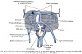
File:Gray0475.jpg ==Human Embryo Liver and Associated Veins== Human embryo, 24 or 25 days old (liver ventral surface view) Figure after His.(2,042 × 1,363 (350 KB)) - 12:27, 4 March 2015
File:Hubrecht Homo32 fig02.jpg Based on the external appearance and limb development this embryo appears to be Stage 20. ...ose, external acoustic meatus, auricle of external ear, arm, elbow, wrist, liver prominence, digital rays(914 × 1,200 (127 KB)) - 06:11, 30 September 2013
File:Hubrecht Homo32 fig01.jpg Based on the external appearance and limb development this embryo appears to be Stage 20. ...ose, external acoustic meatus, auricle of external ear, arm, elbow, wrist, liver prominence, digital rays(872 × 1,200 (159 KB)) - 06:11, 30 September 2013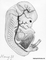
File:Hubrecht Homo32 fig03.jpg ...rom the Hubrecht catalogue page. Based on the external appearance and limb development this embryo appears to be Stage 20. ...ose, external acoustic meatus, auricle of external ear, arm, elbow, wrist, liver prominence, digital rays(618 × 800 (114 KB)) - 06:10, 30 September 2013


