Search results
From Embryology
Page title matches

File:O'Rahilly1957 plate01.jpg {{Ref-O'Rahilly1957}}(2,105 × 2,990 (1.22 MB)) - 17:01, 5 June 2016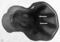
File:O'Rahilly1957 fig01.jpg {{Ref-O'Rahilly1957}}(1,606 × 1,129 (262 KB)) - 17:07, 5 June 2016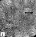
File:O'Rahilly1957 fig02.jpg {{Ref-O'Rahilly1957}}(818 × 826 (285 KB)) - 17:15, 5 June 2016
File:Ronan O'Rahilly.jpg ==Ronan O'Rahilly== Ronan O'Rahilly September 13, 1921 - June 24, 2018.(433 × 632 (14 KB)) - 09:34, 29 December 2018
File:O'Rahilly1956 plate01.jpg (1,599 × 2,032 (293 KB)) - 15:02, 2 July 2018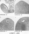
File:O'Rahilly1956 plate02.jpg (1,579 × 1,885 (367 KB)) - 15:01, 2 July 2018
File:O'Connor1940 plate01.jpg {{Ref-O'Connor1940}}(1,280 × 1,503 (268 KB)) - 12:32, 14 November 2018
File:Ronan O'Rahilly 1987.jpg [[Embryology History - Ronan O'Rahilly|Ronan O'Rahilly]] (1987) at the Carnegie labs.(600 × 575 (81 KB)) - 22:08, 23 January 2019


Page text matches
File:Ronan O'Rahilly.jpg ==Ronan O'Rahilly== Ronan O'Rahilly September 13, 1921 - June 24, 2018.(433 × 632 (14 KB)) - 09:34, 29 December 2018File:Ronan O'Rahilly 1987.jpg [[Embryology History - Ronan O'Rahilly|Ronan O'Rahilly]] (1987) at the Carnegie labs.(600 × 575 (81 KB)) - 22:08, 23 January 2019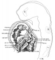
File:Wen1928-Fig14.jpg ...o show the endocardium. Drawn from a model by [[:File:Osborne Heard.jpg|O. O. Heard]] on the basis of a projection reconstruction. X 100.(1,111 × 1,200 (216 KB)) - 15:33, 21 April 2016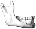
File:Gray0184.jpg ...vertical in direction, the angle measuring from 110<sup>o</sup> to 120<sup>o</sup>.(600 × 491 (45 KB)) - 10:10, 14 September 2012
File:Foster103.jpg o. place of future area vasculosa ; rf. medullary groove ; pr. primitive stre In the region o. a layer of mesoblast has already grown ; there are however as yet no signs(896 × 905 (87 KB)) - 18:04, 12 January 2011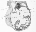
File:Abbott 19.jpg ...ifid apex seen at this stage and also the presence of two openings (O' and O") in the primitive auricular septum.(880 × 791 (171 KB)) - 10:22, 16 September 2020
File:Osborne Heard.jpg ==Osborne O. Heard== Osborne O. Heard (1891–1983) produced model reconstructions for the Carnegie Instit(800 × 571 (93 KB)) - 21:25, 31 January 2019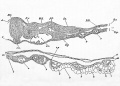
File:Foster034.jpg ...ity, c. h. notochord. a. o. dorsal aorta, v. blood-vessels of the yolksac, o.p. line of junction between opaque and pellucid areas ; w. palisade-like yo(1,003 × 717 (153 KB)) - 08:47, 11 January 2011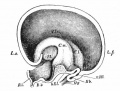
File:Bailey437.jpg .... y occipital lobe; Og. } olfactory nerve; R. i., recessus infundibuli; R. o., recessus (prae-?) opticus; St., stalk of hemisphere (strio-thalamic junct(526 × 397 (50 KB)) - 23:04, 22 February 2011
File:Otto Grosser.jpg {{Online Editor}} - The template {{Grosser, O.}} links to this page. {{Grosser, O.}} Anatomie und Entwicklungsgeschichte der Eihäute und der Placenta. Wien,(400 × 590 (19 KB)) - 11:49, 29 July 2019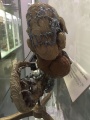
File:Model Carnegie Embryo 588-1.jpg [[:File:Osborne Heard.jpg|Osborne O Heard]] model of vascular system of [[:Category:Carnegie Embryo 588|Embryo ...y:Carnegie Embryo 588|Embryo No. 588]] | [[:File:Osborne Heard.jpg|Osborne O Heard]] | [[Carnegie Collection]](750 × 1,000 (134 KB)) - 00:02, 21 April 2016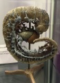
File:Model Carnegie Embryo 588.jpg [[:File:Osborne Heard.jpg|Osborne O Heard]] model of vascular system of [[:Category:Carnegie Embryo 588|Embryo ...y:Carnegie Embryo 588|Embryo No. 588]] | [[:File:Osborne Heard.jpg|Osborne O Heard]] | [[Carnegie Collection]](800 × 1,107 (191 KB)) - 00:02, 21 April 2016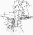
File:Fleming1927-fig01.jpg Drawing of upper part of block while in xylol. b = bladder, o = ovary, r = rectum, t=Fallopian tube, ur = ureter, ut = uterus, v = blood(953 × 1,000 (167 KB)) - 13:39, 15 November 2015
File:Bailey491.jpg O. Schultze.(728 × 473 (56 KB)) - 09:04, 3 February 2011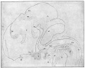
File:Bailey432.jpg ...rve; S., fillet; St., stria medullaris thalami; T., thalamic radiation; T. o., tractus opticus; V, Gasserian ganglion; VII, facial nerve and geniculate(794 × 635 (92 KB)) - 01:21, 14 February 2011
File:AnsonKarabinMartin1939 fig07-12.jpg ...tibule; vestib. orif. or v. o. fiss, vestibular orifice of fissula, and v. o. foss., vestibular orifice of fossula.(1,280 × 1,880 (444 KB)) - 08:39, 22 October 2017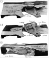
File:AnsonKarabinMartin1939 fig13-15.jpg ...tibule; vestib. orif. or v. o. fiss, vestibular orifice of fissula, and v. o. foss., vestibular orifice of fossula.(1,280 × 1,519 (171 KB)) - 08:46, 22 October 2017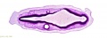
File:Stage 13 image 002.jpg '''A2 and A3:''' [[O#otocyst|Otocyst]] , otic capsule, [[R#rhombencephalon|rhombencephalon]] an '''A2:''' [[O#otocyst|Otocyst]] (right). Apex of [[O#otocyst|otocyst]] (origin of L endolymphatic sac)(1,000 × 359 (52 KB)) - 18:18, 19 August 2010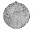
File:Minot1897 fig003.jpg Around Each Pro-nucleus is shown the Aster. — (''After O. Hertwig'')(625 × 543 (83 KB)) - 08:07, 5 April 2014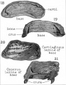
File:AnsonKarabinMartin1939 fig18-21.jpg ...tibule; vestib. orif. or v. o. fiss, vestibular orifice of fissula, and v. o. foss., vestibular orifice of fossula.(1,280 × 1,643 (180 KB)) - 09:01, 22 October 2017


