Search results
From Embryology
Page title matches

File:Stage8 nodal cilia.jpg (450 × 321 (50 KB)) - 12:32, 6 September 2014
File:Nodal cilia 001.mp4 (2.44 MB) - 09:16, 7 March 2013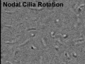
File:Nodal-cilia-001-icon.jpg ...al. (2005) De Novo Formation of Left–Right Asymmetry by Posterior Tilt of Nodal Cilia. PLoS Biol 3(8): e268 [http://www.ncbi.nlm.nih.gov/pubmed/16035921?do ...al. (2005) De Novo Formation of Left–Right Asymmetry by Posterior Tilt of Nodal Cilia. PLoS Biol 3(8): e268. doi:10.1371/journal.pbio.0030268(320 × 240 (13 KB)) - 15:03, 14 October 2009
File:Regulation of Nodal-Activin signalling during heart formation.png ====Regulation of Nodal gradient and signalling activity==== ...is mediated by high Nodal production in epiblast cells, timed secretion of Nodal antagonists (cerberus and LEFTY1) by regions of the visceral endoderm, and(696 × 511 (238 KB)) - 12:26, 26 October 2017


Page text matches
File:Regulation of Nodal-Activin signalling during heart formation.png ====Regulation of Nodal gradient and signalling activity==== ...is mediated by high Nodal production in epiblast cells, timed secretion of Nodal antagonists (cerberus and LEFTY1) by regions of the visceral endoderm, and(696 × 511 (238 KB)) - 12:26, 26 October 2017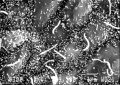
File:Stage8 sem3.jpg ==Scanning EM showing detail of the Nodal Cilia== Human embryo ([[Carnegie stage 8|Stage 8]], day 18) nodal cilia are the long cellular processes, the shorter processes visible on cel(1,000 × 709 (91 KB)) - 23:16, 14 December 2013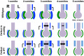
File:Mouse left-right axis 01.jpg ...onse to fluid flow. With reduced expression of its antagonist on the left, Nodal activity and expression (dark blue) increases on the left side and decrease * 2-somite stage wild type embryos, Nodal induce Nodal expression in the L-LPM(1,400 × 937 (172 KB)) - 12:36, 30 April 2017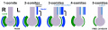
File:Mouse left-right axis 02.jpg Gastrula stage - Initially perinodal crown cells symmetrically express Nodal, Wnt and their antagonist Cerberus like-2 (Cerl2). ...onse to fluid flow. With reduced expression of its antagonist on the left, Nodal activity and expression (dark blue) increases on the left side and decrease(1,313 × 374 (72 KB)) - 17:08, 30 April 2017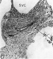
File:Licata1954 fig16b.jpg ...) to show the primordium of the sino-atrial node. Note the position of the nodal artery. Abbreviations: NA, nodal artery; SA, sino-atrial node; Sh, connective-tissue sheath; SVC, superior v(784 × 874 (117 KB)) - 12:04, 5 March 2017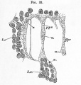
File:Foster033.jpg ...ently become the nuclei of the cells forming the walls of the vessels. The nodal groups are united by protoplasmic processes (p*pr\ also containing nuclei w(654 × 687 (80 KB)) - 08:08, 9 January 2011
File:Embryo left-right asymmetry pathway.jpg * '''a''' - An 8.25-day mouse embryo showing asymmetric Nodal expression (in blue) at the node as well as in the left, but not the right, ...diameters to the left lateral plate mesoderm (LPM). Here it activates the Nodal signaling cascade, ultimately resulting in left-side-specific Pitx2 express(800 × 458 (46 KB)) - 16:30, 12 August 2016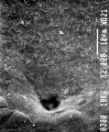
File:Stage8 sem2.jpg ...o ([[Carnegie stage 8|Stage 8]], day 18) showing the neurenteric canal and nodal cilia. The [[:File:Stage8_sem3.jpg|nodal cilia]] are thought to act to "stir" the overlying fluid layer at the time(822 × 1,000 (142 KB)) - 08:55, 23 August 2018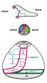
File:Sea urchin ectoderm patterning model 01.jpg ==Changes in identity of ectodermal territories following perturbations of Nodal or BMP signaling and novel model of ectoderm patterning== ...ling in the lateral ectoderm likely contributes to maintain a low level of Nodal and BMP signaling within the presumptive ciliary band region by phosphoryla(300 × 528 (28 KB)) - 17:56, 12 April 2011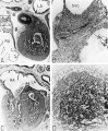
File:Licata1954 fig16.jpg ...ino-atrial node. Note the position of the nodal artery. Abbreviations: NA, nodal artery; SA, sino-atrial node; Sh, connective-tissue sheath; SVC, superior v(1,000 × 1,212 (297 KB)) - 12:20, 5 March 2017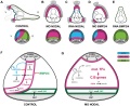
File:Sea urchin ectoderm patterning model.jpg ==Changes in identity of ectodermal territories following perturbations of Nodal or BMP signaling and novel model of ectoderm patterning== (B) Nodal morphant. Most of the ectoderm differentiates into an expanded large ciliar(600 × 490 (74 KB)) - 17:54, 12 April 2011File:Nodal-cilia-001-icon.jpg ...al. (2005) De Novo Formation of Left–Right Asymmetry by Posterior Tilt of Nodal Cilia. PLoS Biol 3(8): e268 [http://www.ncbi.nlm.nih.gov/pubmed/16035921?do ...al. (2005) De Novo Formation of Left–Right Asymmetry by Posterior Tilt of Nodal Cilia. PLoS Biol 3(8): e268. doi:10.1371/journal.pbio.0030268(320 × 240 (13 KB)) - 15:03, 14 October 2009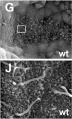
File:Mouse primitive node cilia.jpg Extract from Figure 6: Nodal cilia are present but abnormal in Tbx6 mutant embryos. (A–F) Confocal ima ...re short with bulbous tips in the mutant. (G–L) SEM images of the node and nodal cilia in wt and −/− nodes (G,J) Normal, long filamentous cilia in wild(592 × 981 (130 KB)) - 09:39, 15 April 2010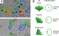
File:Node cilia movement.png ...al. (2005) De Novo Formation of Left–Right Asymmetry by Posterior Tilt of Nodal Cilia. PLoS Biol 3(8): e268 [http://www.ncbi.nlm.nih.gov/pubmed/16035921?do ...al. (2005) De Novo Formation of Left–Right Asymmetry by Posterior Tilt of Nodal Cilia. PLoS Biol 3(8): e268. doi:10.1371/journal.pbio.0030268(600 × 370 (248 KB)) - 15:04, 14 October 2009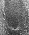
File:Stage8 sem1.jpg ...ge8_sem2.jpg|Image - neurenteric canal]] | [[:File:Stage8_sem3.jpg|Image - nodal cilia]] | {{notochord}} | [[Carnegie stage 8|Stage 8]] | [[Gastrulation]](828 × 1,000 (109 KB)) - 08:53, 23 August 2018
File:Lymphatic-system-overview.jpg # '''Diffuse''' - (extra-nodal tissue) nodules, Mucosal Associated Lymphoid Tissues (MALT)(800 × 755 (118 KB)) - 12:48, 24 February 2016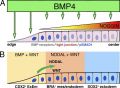
File:Gastrulation molecular factors01.jpg (B) Adjacent to the colony edge, BMP-dependent {{NODAL}} and {{WNT}} expression establishes a {{mesoderm}}/{{endoderm}} cell popul :'''Links:''' {{gastrulation}} | {{BMP}} | {{WNT}} | {{NOGGIN}} | {{NODAL}}(750 × 549 (105 KB)) - 10:50, 29 June 2020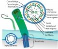
File:Cilium cartoon.jpg :'''Links:''' [[:File:Stage8_sem3.jpg|Human nodal cilia SEM image]](800 × 682 (114 KB)) - 15:46, 27 March 2019
File:Zebrafish ectodermal patterning model.jpg :'''Links:''' {{gastrulation}} | {{BMP}} | {{WNT}} | {{NOGGIN}} | {{NODAL}}(1,280 × 662 (80 KB)) - 10:43, 29 June 2020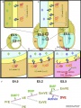
File:Mouse distal visceral endoderm 01.jpg a, Activin; b, BMP; Epi, epiblast; ICM, inner cell mass; l, Lefty1; n, Nodal; PE, parietal endoderm; PrE, primitive endoderm; s1p, phosphorylated Smad1/(959 × 1,280 (208 KB)) - 13:16, 3 May 2013


