Search results
From Embryology
File:Stage 22 image 322.jpg ===Ventricular System=== ===Neural System===(1,180 × 999 (254 KB)) - 11:38, 14 June 2016
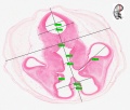
File:Stage22 HPA2L.jpg * Small region from entire cross-section of the [[:Image:Stage22_HPA1L.jpg|stage 22 embryo head]] shown in hi {| class="wikitable mw-collapsible mw-collapsed"(603 × 399 (82 KB)) - 11:51, 14 May 2017
File:Human- ventricular system cartoon.jpg ==Mammalian Ventricular System== ..., and in contact with the cerebrospinal fluid (CSF). The complexity of the system suggests that CSF functions are not limited to metabolic support of the bra(600 × 638 (47 KB)) - 08:26, 24 February 2020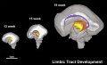
File:Brain tract development 06.jpg ==Brain Tract Development - Limbic Tracts== ...lobe involved in control of emotional expression. Components of the limbic system communicate by the cingulum, white matter fibers projecting from the cingul(1,000 × 605 (35 KB)) - 09:29, 20 November 2019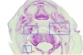
File:Stage 22 image 159.jpg ==Head - Neural, Sensory, Palate, Skull== ...ner Ear Development]] | [[Musculoskeletal_System_-_Skull_Development|Skull Development]](1,000 × 665 (118 KB)) - 11:59, 28 April 2011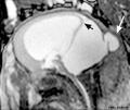
File:Dandy Walker malformation MRI 01.jpg | width=500px|Sagittal b-FFE image of a fetal brain at 26 W shows a markedly enlarged posterior fossa * '''white arrow''' - posterior cephalocele (posterior encephalocele) a neural tube defect. Herniation of the brain and/or meninges through a defect in th(600 × 506 (37 KB)) - 10:29, 1 March 2015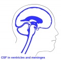
File:Csf cartoon2.jpg Cerebrospinal fluid (CSF) is found in the brain ventricular spaces, spinal cord central canal and the subarachnoid space (between arach Brain four ventricles and several foramina (openings that connect ventricular spaces)(600 × 588 (44 KB)) - 17:18, 26 May 2017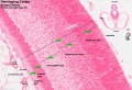
File:Stage 22 image 217.jpg | Human embryo, [[Week 8]], [[Carnegie stage 22]] section from the neural tube at the level of the developing cortex. Inset (upper right) shows secti ...layered structure of the cortex. Layers are named according to the nervous system revised terminology (1970)<ref name=PMID5414696><pubmed>5414696</pubmed></r(1,200 × 820 (323 KB)) - 10:40, 27 May 2017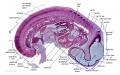
File:Stage 13 image 098.jpg ...:File:Stage 13 image 097.jpg|Previous]] | Last Image | [[Carnegie_stage_13_-_serial_sections#Labeled_Sections|All sections]] ===Cardiovascular System===(1,000 × 623 (144 KB)) - 18:07, 23 August 2010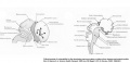
File:CNS later development.jpg '''CNS Later Development''' (E) The lateral view shows the migratory paths from the more central ventricular zone and gradients maturation of the neocortex (see arrows).(961 × 462 (59 KB)) - 22:44, 7 April 2010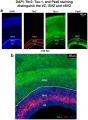
File:Somatosensory cortex of E20 rat.jpeg ...SVZ) and outer SVZ (oSVZ) are identified through a combination of DAPI, Tau-1, Tbr2 and Pax6 staining. Copyright: © 2012 Martínez-Cerdeño et al. This is an open-access article distributed under the terms of the Creative Commons Attributi(2,003 × 2,722 (1.29 MB)) - 12:04, 9 November 2014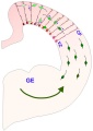
File:Interneuron-radial glial interactions.jpg ==Interneuron-radial glial interactions in the developing cerebral cortex== ...they ascend to the cortical plate (CP) or descend in the direction of the ventricular zone (VZ).(510 × 720 (33 KB)) - 13:30, 12 October 2015
File:Adult human hypothalamus 02.jpg * '''b''' - 3D overview of hypothalamic nuclei, constructed from a 3D high-field MRI data set; left, frontal view; intermediate, lateral view; right, m * Vf - ventricular foramen(1,151 × 343 (100 KB)) - 07:51, 16 May 2014
File:Thyng1914 plate4b.jpg ==Plate 4. Heart, Right Venous System and Left Placental Vein== ...nstruction to show the right atrium and ventricle of the heart; the venous system of the right side; and the left umbilical vein of a 17.8 human embryo (H. E(1,142 × 1,500 (336 KB)) - 14:21, 18 May 2014


