Search results
From Embryology
Page title matches
File:Ultrasound - Hypoplastic left heart syndrome 03.jpg ==Ultrasound Hypoplastic Left Heart Syndrome== LA - left atria(874 × 612 (51 KB)) - 16:22, 22 June 2016
File:Ultrasound - Hypoplastic left heart syndrome 04.jpg ==Ultrasound Hypoplastic Left Heart Syndrome== ...ation shows flow from atrium into ventricle on the right side but not the left.(919 × 618 (68 KB)) - 16:26, 22 June 2016
File:US Hypoplastic Left Heart Syndrome GA19week.mp4 (3.94 MB) - 16:36, 22 June 2016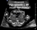
File:Hypoplastic left heart syndrome movie icon.jpg (200 × 165 (10 KB)) - 16:50, 22 June 2016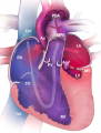
File:Hypoplastic Left Heart Syndrome (HLHS).png ...yndrome (HLHS).RA. Right Atrium. RV. Right Ventricle. LA. Left Atrium. LV. Left Ventricle. SVC. Superior Vena Cava. IVC. Inferior Vena Cava. MPA. Main Pulm Centers for Disease Control and Prevention. (2016). Congenital Heart Defects. Retrieved from https://www.cdc.gov/ncbddd/heartdefects/hlhs.html(351 × 464 (254 KB)) - 12:34, 26 October 2017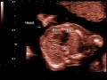
File:Ultrasound - Hypoplastic left heart syndrome 01.jpg ==Ultrasound Hypoplastic Left Heart Syndrome== LA - left atria(800 × 600 (53 KB)) - 16:04, 22 June 2016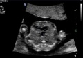
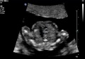
File:Ultrasound - Hypoplastic left heart syndrome 02.jpg ==Ultrasound Hypoplastic Left Heart Syndrome== The four chamber heart view shows a small left atrium (LA) and tiny left ventricle (LV).(890 × 626 (54 KB)) - 16:22, 22 June 2016


.png)


Page text matches
File:Ultrasound - Hypoplastic left heart syndrome 01.jpg ==Ultrasound Hypoplastic Left Heart Syndrome== LA - left atria(800 × 600 (53 KB)) - 16:04, 22 June 2016File:Ultrasound - Hypoplastic left heart syndrome 02.jpg ==Ultrasound Hypoplastic Left Heart Syndrome== The four chamber heart view shows a small left atrium (LA) and tiny left ventricle (LV).(890 × 626 (54 KB)) - 16:22, 22 June 2016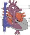
File:Hypoplastic Left Heart.jpg ==Heart Abnormality - Hypoplastic Left Heart== * 4-8% of Congenital Heart Disease(297 × 350 (17 KB)) - 10:33, 19 February 2017File:Ultrasound - Hypoplastic left heart syndrome 04.jpg ==Ultrasound Hypoplastic Left Heart Syndrome== ...ation shows flow from atrium into ventricle on the right side but not the left.(919 × 618 (68 KB)) - 16:26, 22 June 2016File:Ultrasound - Hypoplastic left heart syndrome 03.jpg ==Ultrasound Hypoplastic Left Heart Syndrome== LA - left atria(874 × 612 (51 KB)) - 16:22, 22 June 2016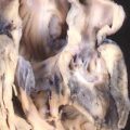
File:Anderson2016-fig44a.jpg ==Fig. 44a. Hypoplastic Left Heart Syndrome== ...ral stenosis with aortic atresia. The right hand panel shows the slit-like left ventricle, without fibroelastosis, found when there is mitral atresia and a(800 × 800 (83 KB)) - 12:25, 19 February 2017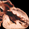
File:Anderson2016-fig44b.jpg ==Fig. 44b. Hypoplastic Left Heart Syndrome== ...ral stenosis with aortic atresia. The right hand panel shows the slit-like left ventricle, without fibroelastosis, found when there is mitral atresia and a(800 × 800 (76 KB)) - 12:26, 19 February 2017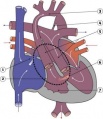
File:Functional Hypoplastic Left Heart.jpg Functional Hypoplastic Left Heart * 4-8% of Congenital Heart Disease(302 × 350 (18 KB)) - 02:28, 27 March 2010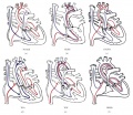
File:Normal fetal blood flow and Tetralogy of Fallot.jpg ...tion of the Great Arteries. (e) tetralogy of fallot. (f) hypoplastic right heart. Red arrows: oxygenated blood; blue arrows: deoxygenated blood.(628 × 543 (200 KB)) - 17:21, 28 October 2011File:Hypoplastic Left Heart Syndrome (HLHS).png ...yndrome (HLHS).RA. Right Atrium. RV. Right Ventricle. LA. Left Atrium. LV. Left Ventricle. SVC. Superior Vena Cava. IVC. Inferior Vena Cava. MPA. Main Pulm Centers for Disease Control and Prevention. (2016). Congenital Heart Defects. Retrieved from https://www.cdc.gov/ncbddd/heartdefects/hlhs.html(351 × 464 (254 KB)) - 12:34, 26 October 2017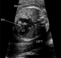
File:ZPulmonary Atresia.jpg ...tricle is greater in size than the right ventricle. The right ventricle is hypoplastic, and the right ventricular cavity is small.(653 × 618 (85 KB)) - 14:29, 19 August 2014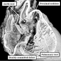
File:Anderson2016-fig46b.jpg ==Fig. 46b. Mouse E14.5 Heart== ...ght ventricle. This can occur when the proximal cushions themselves remain hypoplastic, and the interventricular communication remains doubly committed, as shown(800 × 800 (120 KB)) - 22:39, 16 February 2017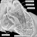
File:Anderson2016-fig46a.jpg ==Fig. 46a. Mouse E12.5 Heart== ...ght ventricle. This can occur when the proximal cushions themselves remain hypoplastic, and the interventricular communication remains doubly committed, as shown(800 × 800 (100 KB)) - 10:36, 6 June 2017


