Search results
From Embryology
Page title matches
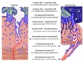
File:Placenta humans and guinea-pig cartoon.jpg (1,200 × 889 (562 KB)) - 22:01, 25 November 2013

Page text matches
File:Guineapig icon.jpg (240 × 180 (7 KB)) - 23:51, 17 January 2013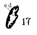
File:Anson-1934 fig17.jpg ...cings of endolymphatic duct and adjacent area of membranous labyrinth from Guinea-pig (18.5 mm)== Guinea-pig: 17, 18.5 mm.(198 × 189 (6 KB)) - 09:31, 3 February 2017
File:Skeletal muscle histology 004.jpg (1,280 × 1,024 (242 KB)) - 15:33, 6 March 2012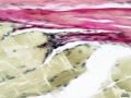
File:Skeletal muscle histology 444.jpg (934 × 701 (125 KB)) - 15:34, 6 March 2012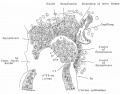
File:Bandler1983 fig06.jpg ==Fig. 6. Further stage of embedding of guinea-pig's ovum==(1,000 × 787 (168 KB)) - 23:07, 11 September 2018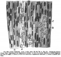
File:Keibel Mall 332.jpg (851 × 800 (142 KB)) - 20:58, 9 February 2014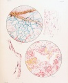
File:Wislocki1920 plate 2.jpg Fig. 7. Kidney of guinea-pig fetus measuring 36 mm., showing trypan-blue in the convoluted tubules 72 ho Fig. 8. Section of the umbilical cord of a guinea-pig fetus measuring 42 mm., showing vitally stained mucoid connective-tissue ce(988 × 1,200 (245 KB)) - 10:47, 16 June 2013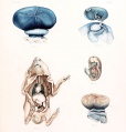
File:Wislocki1920 plate 1.jpg Fig. 2. Gross appearance of a guinea-pig fetus with the amnion opened 36 hours after injection of trypan-blue into t Fig. 3. Guinea-pig fetus, nearly full term, after injection of potassium ferrocyanide and iron(1,145 × 1,200 (173 KB)) - 10:48, 16 June 2013
File:Bandler1983 fig01.jpg ==Fig. 1. Half-schematic section of the uterine horn of a guinea-pig==(1,000 × 903 (179 KB)) - 22:24, 11 September 2018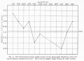
File:Stockard Papanicolaou1917 figA.jpg ==Length in Days of Guinea-pig Diestrous Cycle during the Year== ...d George N. Papanicolaou. The existence of a typical oestrous cycle in the guinea-pig— with a study of its histological and physiological changes. The American(1,382 × 1,000 (143 KB)) - 21:27, 4 November 2013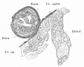
File:Bandler1983 fig03a.jpg ==Fig. 3a. Ovum of guinea-pig in its first adhesion to the uterine epithelium==(900 × 720 (157 KB)) - 22:44, 11 September 2018
File:Wislocki1920 plate 3.jpg Fig. 12. Section through placenta of guinea-pig fetus measuring 40 mm., showing a vitally stained macrophage 26 hours after(975 × 1,200 (219 KB)) - 10:46, 16 June 2013
File:Marshall Edwards.jpg (500 × 370 (48 KB)) - 11:24, 24 December 2019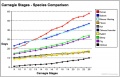
File:Carnegie stages species comparison.jpg ...f>Harman, MT and Dobrovolny, P. he development of the external form of the guinea-pig (Cavia cobaya) between the ages of 11 days and 20 days of gestation. J. of(800 × 514 (85 KB)) - 18:16, 3 June 2011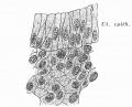
File:Bandler1983 fig02.jpg ...of the compact zone with the epithelial lining of the uterine lumen of the guinea-pig==(800 × 648 (134 KB)) - 22:31, 11 September 2018
File:Bandler1983 fig04.jpg ==Fig. 4. Ovum of guinea-pig partially embedded==(953 × 638 (175 KB)) - 22:50, 11 September 2018
File:Bandler1983 fig03.jpg ==Fig. 3. Ovum of guinea-pig free in the uterine cavity==(700 × 578 (88 KB)) - 22:37, 11 September 2018File:Placenta humans and guinea-pig cartoon.jpg (1,200 × 889 (562 KB)) - 22:01, 25 November 2013
File:Placental trophospongium.jpg (567 × 344 (94 KB)) - 13:35, 18 July 2019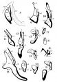
File:Anson-1934 fig08-21.jpg Guinea-pig: 17, 18.5 mm. File:Anson-1934 fig17.jpg|17 Guinea-pig 18.5 mm(1,000 × 1,435 (178 KB)) - 09:40, 3 February 2017


