Search results
From Embryology
Page title matches
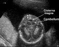
File:Second Trimester Cerebellum.jpeg =Second Trimester Cerebellum= Inferior image of a fetal cerebellum at second trimester(475 × 385 (26 KB)) - 14:47, 16 July 2018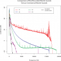
File:Third trimester sounds 01.png [[Category:Third Trimester}}(1,280 × 1,289 (335 KB)) - 09:44, 12 April 2019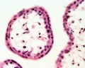
File:Placenta- first trimester histology x40.jpg First trimester human x40 {{HE}}(1,000 × 800 (124 KB)) - 13:50, 23 February 2013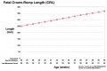
File:Third trimester Crown-Rump Length graph.jpg ==Third trimester Crown-Rump Length Graph== [[Category:Human]] [[Category:Fetal]] [[Category:Third Trimester]] [[Category:Graph]](972 × 648 (59 KB)) - 10:06, 15 June 2016
File:Third trimester maternal womb sounds 01.mp3 :'''Links:''' [[Audio]] | [[Second Trimester]] | [[Third Trimester]] [[Category:Third Trimester]][[Category:Audio]](6.93 MB) - 09:49, 12 April 2019


Page text matches
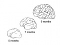
File:Dev anat 01.jpg ==Third Trimester Brain Growth== ...external lateral (left) view of the brain growth that occurs in the third trimester of human development.(500 × 375 (25 KB)) - 09:38, 18 March 2012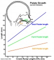
File:Fetal palate growth graph.jpg Growth of the fetal palate and its components during the second trimester. :[[Palate Development|'''Links''']]: [[Palate Development]] | [[Second Trimester]](681 × 757 (77 KB)) - 12:35, 7 May 2016File:Second Trimester Cerebellum.jpeg =Second Trimester Cerebellum= Inferior image of a fetal cerebellum at second trimester(475 × 385 (26 KB)) - 14:47, 16 July 2018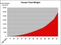
File:Fetal weight change.jpg ...ght change (increase) is greatest in the final weeks of development (third trimester). :'''Links:''' [[Third Trimester]] | [[Birth]](600 × 444 (34 KB)) - 05:08, 29 November 2011File:Third trimester maternal womb sounds 01.mp3 :'''Links:''' [[Audio]] | [[Second Trimester]] | [[Third Trimester]] [[Category:Third Trimester]][[Category:Audio]](6.93 MB) - 09:49, 12 April 2019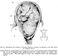
File:Frazer006 bw600.jpg ==Fetus during Third Trimester in Uterus compared to Non-pregnant Uterus== [[Category:Historic Embryology]] [[Category:Human Fetus]] [[Category:Third Trimester]] [[Category:Cartoon]](600 × 575 (47 KB)) - 17:30, 1 June 2013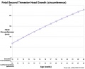
File:Fetal head growth circumference graph02.jpg ==Fetal Second Trimester Head Growth Circumference== ...[[Category:Fetal]] [[Category:Head]] [[Category:Graph]] [[Category:Second Trimester]](800 × 650 (44 KB)) - 14:07, 26 May 2019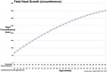
File:Fetal head growth circumference graph01.jpg ...y:Head]] [[Category:Graph]] [[Category:Second Trimester]] [[Category:Third Trimester]](905 × 613 (58 KB)) - 12:05, 15 April 2020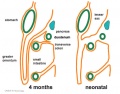
File:Greater-omentum.jpg ...The figure shows a lateral view of this process comparing the early second trimester arrangement with the newborn structure. ...] [[Category:Gastrointestinal Tract]] [[Category:Fetal]] [[Category:Second Trimester]] [[Category:Neonatal]] [[Category:Cartoon]](537 × 419 (48 KB)) - 00:16, 3 May 2011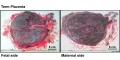
File:Placenta term anatomy 01.jpg ...nta Development|'''Placenta Links''']]: [[Placenta Development]] | [[Third Trimester]] | [[Birth]] [[Category:Human]] [[Category:Placenta]] [[Category:Third Trimester]](1,200 × 600 (115 KB)) - 11:04, 8 May 2018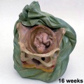
File:Galletti1770 week 16.jpg [[Category:Second Trimester]](450 × 450 (32 KB)) - 08:00, 27 May 2010File:Third trimester Crown-Rump Length graph.jpg ==Third trimester Crown-Rump Length Graph== [[Category:Human]] [[Category:Fetal]] [[Category:Third Trimester]] [[Category:Graph]](972 × 648 (59 KB)) - 10:06, 15 June 2016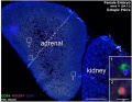
File:Fetal adrenal ectopic germ cells 04.jpg ==Human Fetal Adrenal Ectopic Germ cells (first trimester) == Histological sections of human adrenal (and adjacent kidney) in first trimester female week 11 ({{GA}} week 13).(899 × 700 (147 KB)) - 12:11, 18 July 2018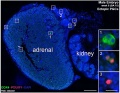
File:Fetal adrenal ectopic germ cells 03.jpg ==Human Fetal Adrenal Ectopic Germ cells (first trimester) == Histological sections of human adrenals (and adjacent kidney) in first trimester male week 8 ({{GA}} week 10)(899 × 700 (159 KB)) - 12:12, 18 July 2018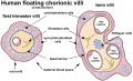
File:Human placental villi cartoon 01.jpg ...{{placental villi}} between early ({{first trimester}}) and late ({{third trimester}}) placental development. # {{Hofbauer cells}} are abundant in villi in both first and second trimesters (though only shown in the term villi). Later {{Hofbau(1,084 × 663 (142 KB)) - 09:31, 26 March 2019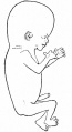
File:Bailey094.jpg [[Category:Human]] [[Category:Fetal]] [[Category:Second Trimester]](376 × 689 (27 KB)) - 00:30, 14 February 2011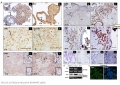
File:Expression of CD82 in human placental villi and cell lines.JPG ...nvasive extravillous trophoblast cell line derived from immortalized first trimester trophoblast; B6Tert: immortalized cytotrophoblast cell line; JEG-3: human c(612 × 432 (75 KB)) - 08:42, 8 August 2012
File:Fetal adrenal ectopic germ cells 02.jpg ==Human Fetal Adrenal Ectopic Germ cells (first trimester) == Histological sections of human adrenals (and adjacent kidney) in first trimester male week 8 ({{GA}} week 10) and female week 11 ({{GA}} week 13).(1,086 × 446 (124 KB)) - 12:11, 18 July 2018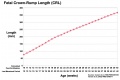
File:Fetal length change.jpg ...s a constant and continual growth in fetal length through second and third trimester. ...:Human]] [[Category:Fetal]] [[Category:Second Trimester]] [[Category:Third Trimester]] [[Category:Graph]](972 × 648 (72 KB)) - 13:47, 26 May 2019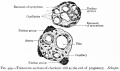
File:Bailey499.jpg [[Category:Human]] [[Category:Placenta]] [[Category:Birth]] [[Category:Third Trimester]](805 × 477 (65 KB)) - 08:03, 2 February 2011


