Search results
From Embryology

File:Smith1957 fig01.jpg ==Fig. 1. Vogt’s classification of atresia of the esophagus== Type I, atresia of entire esophagus.(1,000 × 583 (73 KB)) - 11:13, 24 August 2016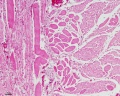
File:Smooth muscle histology 006.jpg * human esophagus(1,280 × 1,024 (481 KB)) - 14:21, 23 February 2013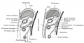
File:Gray0990.jpg ...mall sac lying within the thorax on the right side of the lower end of the esophagus. The anterior layer of the transverse mesocolon is at first distinct from t(800 × 407 (60 KB)) - 15:56, 28 April 2011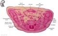
File:Embryo stage 22 C7L.jpg * relative postion of esophagus and trachea(619 × 389 (67 KB)) - 11:33, 31 May 2010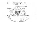
File:Lisser1911 fig01.jpg ...cricoarytacnoideus posterior. V. C., vertebral column; Esoph. L., Lumen of esophagus; constr., constrictor muscle; cri. post, M. cricoarytaenoide'uspostcior; n.(1,119 × 900 (92 KB)) - 14:50, 12 June 2016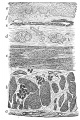
File:Gray1033.jpg ==Fig. 1033. Section of the Human Esophagus==(399 × 600 (96 KB)) - 12:09, 11 May 2014
File:Human week 10 fetus 08.jpg ..., epiglottis, arytenoid cartilage, arytænoideus muscle, cricoid cartilage, esophagus, trachea, vertebra(1,200 × 900 (323 KB)) - 21:03, 8 October 2015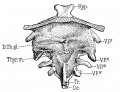
File:Sudler1902-fig05.jpg D. Th. g1., ductus thyreoglossus; Hyp., hypophysis; 0e., (esophagus; Tr., trachea; V. P.’ V. P.’’, V. P./” and V. PJV, flrst, second,(1,000 × 769 (182 KB)) - 15:37, 24 May 2017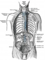
File:Gray0621.jpg ...on of the seventh rib with its cartilage; (b) middle, to the glands on the esophagus and to those around the termination of the inferior vena cava; and (c) post ...and lateral aortic glands and to the glands on the terminal portion of the esophagus.(599 × 800 (131 KB)) - 13:00, 15 February 2013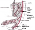
File:GIT blood supply.jpg # '''Foregut''' - celiac artery (Adult: pharynx, esophagus, stomach, upper duodenum, respiratory tract, liver, gallbladder pancreas)(568 × 500 (47 KB)) - 12:48, 17 April 2019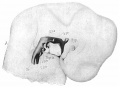
File:Sudler1902-fig04.jpg Hyp., hypophysis; N., outline of the side of the nasal depression; 0e., (esophagus; Thyr. m., median thyroid rudiment; Tr., trachea; V. P.<sup>I</sup>, V. P.<(1,000 × 724 (104 KB)) - 09:51, 22 September 2015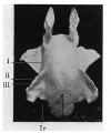
File:Dickie1914 fig06.jpg ...where it again widens, and behind which it tapers away gradually into the (esophagus, the lumen of which is uniformly narrow. The cavity as a whole is curved on(504 × 607 (57 KB)) - 13:17, 20 August 2018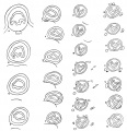
File:Blaisdell1914 fig21.jpg ...e termination of the pharynx; B, through the transitional point; C and D, (esophagus slightly separated from the larynx; E to J, oesophagus contiguous to the tr(1,000 × 1,037 (173 KB)) - 12:33, 14 January 2017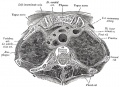
File:Gray0967.jpg ...vein; the vagus, cardiac, phrenic, and left recurrent nerves; the trachea, esophagus, and thoracic duct; the remains of the thymus, and some lymph glands.(700 × 506 (109 KB)) - 03:45, 17 August 2012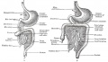
File:Gray0987.jpg ...mall sac lying within the thorax on the right side of the lower end of the esophagus. The anterior layer of the transverse mesocolon is at first distinct from t(1,000 × 579 (114 KB)) - 15:02, 28 April 2011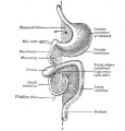
File:Gray0987a.jpg ...mall sac lying within the thorax on the right side of the lower end of the esophagus. The anterior layer of the transverse mesocolon is at first distinct from t(554 × 579 (50 KB)) - 14:02, 21 August 2018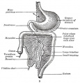
File:Gray0987b.jpg ...mall sac lying within the thorax on the right side of the lower end of the esophagus. The anterior layer of the transverse mesocolon is at first distinct from t(554 × 579 (67 KB)) - 10:13, 26 May 2020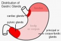
File:Stomach gastric gland distribution.jpg ** similar to the cardiac glands of the esophagus(500 × 334 (28 KB)) - 12:51, 5 April 2013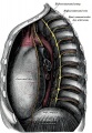
File:Gray0969.jpg ...azygos and the two hemiazygos veins, the vagus and splanchnic nerves, the esophagus, the thoracic duct, and some lymph glands.(621 × 900 (241 KB)) - 18:45, 24 February 2019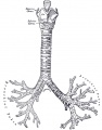
File:Gray0961.jpg * '''Posteriorly''' it is in contact with the esophagus. * passes beneath the aortic arch, crosses in front of the esophagus, the thoracic duct, and the descending aorta, and has the left pulmonary ar(600 × 769 (66 KB)) - 12:01, 18 May 2014


