Search results
From Embryology
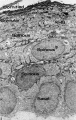
File:Human embryo skin 24 week EGA.jpg ==Keratinized Fetal Epidermis (about 24 week EGA)== Transmission electron micrograph (TEM) of full thickness keratinized epidermis. Note the presence of abundant glycogen in all epidermal layers, the periph(596 × 939 (165 KB)) - 23:51, 28 September 2011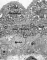
File:Human embryo skin 9-11 week EGA.jpg ...n micrograph (TEM) of the full thickness early stratified fatal epidermis. Epidermis has stratified to three layers. {{Epidermis EM links}}(623 × 804 (176 KB)) - 23:52, 28 September 2011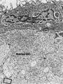
File:Human embryo skin 8-9 week EGA.jpg Transmission electron micrograph (TEM) of embryonic epidermis showing periderm and basal layer. {{Epidermis EM links}}(657 × 872 (188 KB)) - 12:23, 27 March 2019
File:Human embryo skin 8-9 week EGA desmosomes.jpg {{Epidermis EM links}}(800 × 198 (40 KB)) - 12:23, 27 March 2019


