Search results
From Embryology
Page title matches
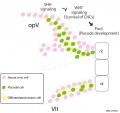
File:Mouse cranial nerve model SHH.jpg ==Mouse Cranial Nerve Model - Sonic Hedgehog== ...om rhombomere 2 (r2) or 4 (r4) interact with placodal cells to develop the cranial nerves.(954 × 900 (74 KB)) - 14:03, 6 May 2018

Page text matches
File:Gray0698.jpg ==Fig. 698. Primary Terminal Nuclei of the Afferent (sensory) Cranial Nerves== * Cranial Nerve 8 (CNVIII) to cochlear nuclei (hearing) and vestibular nuclei (balance)(500 × 518 (47 KB)) - 04:34, 27 April 2014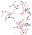
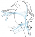
File:Gray0697.jpg ==Fig. 697. Cranial motor nerves brainstem nuclei of origin== * Cranial nerve 7 (CNVII) facial nerve to facial motor nucleus(500 × 540 (49 KB)) - 04:32, 27 April 2014
File:Human week 10 fetus 12.jpg ==Human Female Fetus - Olfactory Nerve (10 week)== ...ed from the nasal placode, the olfactory receptor neurons form an afferent nerve fibre transmitting the [[Sensory - Smell Development|sense of smell]] to ol(1,200 × 900 (349 KB)) - 14:38, 25 May 2016File:Mouse cranial nerve model SHH.jpg ==Mouse Cranial Nerve Model - Sonic Hedgehog== ...om rhombomere 2 (r2) or 4 (r4) interact with placodal cells to develop the cranial nerves.(954 × 900 (74 KB)) - 14:03, 6 May 2018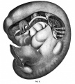
File:Mall1905 fig3.jpg ...reconstructed; the tenth and twelfth cranial nerves and the flrst cervical nerve by [[Embryology History - George Streeter|Dr. Streeter]]. [[Category:Cranial Nerve]](902 × 1,000 (155 KB)) - 12:00, 17 February 2016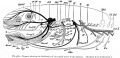
File:Bailey368.jpg ==Fig. 368. Diagram showing the distribution of the cranial nerves in the Amniota== [[Category:Cranial Nerve]](1,074 × 523 (134 KB)) - 08:20, 16 February 2016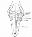
File:Bailey390.jpg ...point of attachment of the acoustic and the sensory root of the trigeminal nerve is shown by dotted circles; the motor nuclei are represented by heavy dots. [[Category:Cranial Nerve]](583 × 667 (50 KB)) - 23:01, 4 September 2014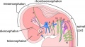
File:Neural - cranial nerves.jpg ==Cranial Nerves== Cranial nerves are associated with the brain, compared to spinal nerves which emerg(800 × 447 (72 KB)) - 21:07, 14 April 2016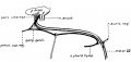
File:Streeter1906 fig07.jpg ==Fig. 7. Facial Nerve Pig Embryo 20 cm== Drawing made from a dissection of the facial nerve ({{CN VII}}) in a 20 cm {{pig}} embryo.(1,254 × 591 (75 KB)) - 17:09, 13 May 2018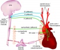
File:Baroreceptor reflex cartoon.jpg :'''Links:''' [[Neural - Cranial Nerve Development|Cranial Nerve Development]] | [[Cardiovascular System Development]] | [[Endocrine - Adren {{Cranial Nerve Links}}(1,200 × 1,012 (182 KB)) - 10:25, 18 December 2018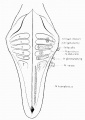
File:Streeter1908 fig01.jpg ...point of attachment of the acoustic and the sensory root of the trigeminal nerve Is shown by dotted circles ; the motor nuclei are represented by heavy dots [[Category:Neural]][[Category:Cranial Nerve]](706 × 1,000 (80 KB)) - 12:57, 22 April 2016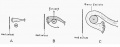
File:Streeter1908 fig02.jpg ==Fig. 2. Diagram illustrating the development of the genu of the facial nerve in the human embryo== The drawings show the right facial nerve and its nucleus of origin, in three stages : the youngest, A. being the 10(1,000 × 391 (39 KB)) - 12:57, 22 April 2016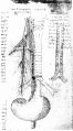
File:McMurrich1930 fig75.jpg ...g. 75. Figure showing the course and distribution of the reversive (vagus) nerve== [[Category:Historic Embryology]][[Category:Neural]][[Category:Cranial Nerve]](1,280 × 2,307 (389 KB)) - 16:16, 21 April 2020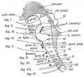
File:Keith1902 fig180.jpg ==Fig. 180. A Diagram to show the Relationship of the Cranial Nerves to the Primitive Segments of the Head== The Segments to which the Cranial Nerves belong.(872 × 800 (141 KB)) - 12:46, 23 May 2016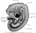
File:Bailey164.jpg ...ronchus; L, liver; K, anlage o kidney; T, thyreoid gland; III-XII, cranial nerve roots ...spinal nerve roots), 1st and 8th cervical, 12th dorsal, 5th lumbar spinal nerve respectively.(928 × 862 (149 KB)) - 14:24, 18 January 2011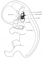
File:Streeter1906 fig02.jpg ...of the seventh is indicated by solid black. The great superflcial petrosal nerve extends from the geniculate to the spheno-palatine ganglion. [[Category:Cranial Nerve]](1,527 × 2,059 (244 KB)) - 16:43, 13 May 2018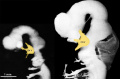
File:Human Stage14-16 CN5-01.jpg ...ee major branches: ophthalmic nerve (V1), maxillary nerve (V2), mandibular nerve (V3) [[Category:Cranial Nerve]](1,028 × 681 (44 KB)) - 13:26, 8 May 2018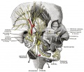
File:Gray0781.jpg ==Fig. 781. Mandibular division of the Trigeminal Nerve== ===Mandibular Nerve===(817 × 700 (156 KB)) - 10:53, 25 June 2018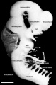
File:Human Stage16 neural02.jpg ...Showing 5 secondary vesicles, 3 brain flexures, eye and otic vesicle, and cranial nerves. | {{Cranial Nerve Table}}(1,352 × 2,048 (286 KB)) - 08:50, 13 October 2017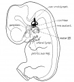
File:Streeter1906 fig01.jpg ...d relations to the brain and the fifth ({{CN V}}) and seventh ({{CN VII}}) cranial nerves. [[Category:Cranial Nerve]](1,193 × 1,318 (167 KB)) - 16:41, 13 May 2018


