Search results
From Embryology

File:Stage12 bf6.jpg ==Human Embryo Carnegie Stage 12== Carnegie Collection Embryo No.5923(350 × 500 (20 KB)) - 13:18, 22 April 2012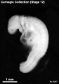
File:Stage12 bf7.jpg ==Human Embryo Carnegie Stage 12== Carnegie Collection Embryo No.6097(350 × 500 (18 KB)) - 13:19, 22 April 2012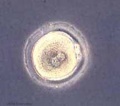
File:Early zygote.jpg :[[Zygote|'''Links''']]: [[Carnegie stage 1]] | [[:File:Early zygote.jpg|Image - Early zygote]] | [[:File:Early zygote ===About Carnegie Stages 1===(500 × 441 (23 KB)) - 12:15, 20 July 2015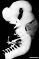
File:Human Stage16 neural03.jpg ==Human Carnegie Stage 16 Neural== ...s been flipped horizontally (to left view) to use in comparison with other stages.(1,352 × 2,048 (245 KB)) - 15:44, 24 May 2017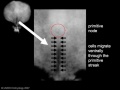
File:Stage7 primitive streak labelled.jpg ==Human Embryo - Carnegie Stages 7== View 1: embryonic disc, showing the epiblast viewed from the amniotic (dorsal) sid(500 × 375 (13 KB)) - 12:49, 9 May 2016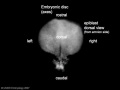
File:Stage7 axes.jpg ==Human Embryo - Carnegie Stages 7== View 1: embryonic disc, showing the epiblast viewed from the amniotic (dorsal) sid(500 × 375 (9 KB)) - 16:51, 4 April 2012
File:Human Stage14-16 CN5-01.jpg ...s been flipped horizontally (to left view) to use in comparison with other stages. ...the right lateral view of the central nervous system of embryo at Carnegie stages {{CS14}} and {{CS16}}.(1,028 × 681 (44 KB)) - 13:26, 8 May 2018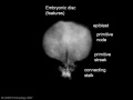
File:Stage7 features.jpg [[Carnegie stage 7]] View 1: embryonic disc, showing the epiblast viewed from the amniotic (dorsal) sid(500 × 375 (9 KB)) - 09:38, 31 July 2018
File:Human embryo head week 6 to 8.jpg ==Lateral view of Embryos between Week 6 to 8 (Carnegie Stage 17 to 23) showing Craniofacial Morphogenesis== * Lateral view of embryos between Carnegie stage 17 and 23 showing craniofacial morphogenesis.(540 × 780 (66 KB)) - 17:12, 23 March 2016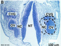
File:Human CS13 otic vesicle 01.jpg ==Human Otic Vesicle Carnegie Stages 13 to 15== Carnegie Stages - {{CS13}}(1,028 × 774 (112 KB)) - 12:34, 6 April 2018
File:Congdon1922-1-16.jpg ==Figs. 1 to 16. Ventral views of aortic-arch system== Showing successive developmental stages. In the earliest stage only the first arch is present, while in the last (a(980 × 1,000 (157 KB)) - 21:30, 6 November 2018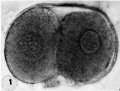
File:Hertig1956 fig01.jpg ==Fig. 1.== ...development; one segmenting egg found in the tube, Streeter Horizon II, [[Carnegie stage 2]], Embryo No. {{CE8698}}, Sequence 12. X 500.(745 × 562 (63 KB)) - 11:21, 16 June 2017
File:Bardeen1906-plate01.jpg Fig. 1. Embryo {{CE2}}, length 7 mm., age about four weeks. ...igures, repeated from this journal (Vol. 1, Plate II), to illustrate early stages in the diiferentiation of the inferior extremity.(1,565 × 2,322 (238 KB)) - 23:06, 23 July 2020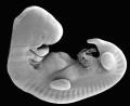
File:Stage14 SEM.jpg Human embryo (Carnegie stage 14) Features: midbrain, nasal placode, lens pit, 1,2,3 pharyngeal arches, fourth ventricle of brain, 1st pharyngeal groove, he(646 × 530 (36 KB)) - 22:47, 3 August 2009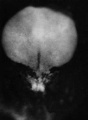
File:Stage7.jpg == Human Embryo - Carnegie stage 7== View 1: embryonic disc, showing the epiblast viewed from the amniotic (dorsal) sid(312 × 427 (7 KB)) - 19:13, 29 August 2013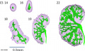
File:Human embryonic renal branching 1.jpg ...t in embryos between week 5 to 8 from the [[Kyoto Collection]] at carnegie stages: {{CS14}}, {{CS16}}, {{CS18}}, {{CS19}} and {{CS22}}. See also [[:File:Huma ...s and urinary collecting system during embryonic periods. Numbers indicate Carnegie stage. Scale bar = 0.5 mm.(1,280 × 779 (236 KB)) - 10:26, 18 January 2019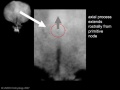
File:Stage7 axial process.jpg ==Human Embryo - Carnegie Stage 7== View 1: embryonic disc, showing the epiblast viewed from the amniotic (dorsal) sid(500 × 375 (11 KB)) - 16:57, 4 April 2012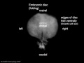
File:Stage7 folding.jpg == Human Embryo - Carnegie stage 7== View 1: embryonic disc, showing the epiblast viewed from the amniotic (dorsal) sid(500 × 375 (10 KB)) - 16:53, 4 April 2012
File:Stage14 human scale.jpg Human embryo (Carnegie stage 14) Features: midbrain, nasal placode, lens pit, 1,2,3 pharyngeal arches, fourth ventricle of brain, 1st pharyngeal groove, he(646 × 530 (40 KB)) - 22:42, 3 August 2009
File:Stage9 dorsal.jpg ==Human Embryo Carnegie Stage 9 (late)== Facts: Week 3, 19 - 21 days, 1.5 - 2.5 mm, Somite Number 1 - 3(287 × 378 (5 KB)) - 07:26, 7 April 2012


